49 results filtered with: Mouse

- Digital Images
- Online
Trichuris muris is a parasitic nematode affecting mice. Following ingestion, T. muris eggs hatch in the large intestine where they develop into adults. The anterior end of the worm burrows into the lining of the gut, leaving the posterior end protruding into the lumen of the gut. The worms mate in this orientation, and the resulting eggs are released in to the gut lumen and shed faecally.
David Goulding, Wellcome Trust Sanger Institute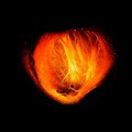
- Digital Images
- Online
Left ventricle, mouse heart, OPT
Laurence Jackson, Centre for Advanced Biomedical Imaging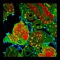
- Digital Images
- Online
Mouse kidney
Kevin Mackenzie, University of Aberdeen
- Digital Images
- Online
Mouse embryo head viewed from the front
Robert Hindges, KCL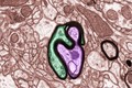
- Digital Images
- Online
Myelinated nerves in a mouse brain, TEM
Mikaela Laine, University of Helsinki
- Digital Images
- Online
Movie of a mouse head in development
NIMR, Francis Crick Institute
- Digital Images
- Online
Dorsal root ganglion neurone from a mouse, LM
Marta Alves Simões, University of Sheffield
- Digital Images
- Online
14.5 dpc mouse forelimb
NIMR, MRC
- Digital Images
- Online
TEM mouse cell, with close-up of inclusion.
David Gregory & Debbie Marshall
- Digital Images
- Online
Nerve in skeletal muscle, showing dystrophin location
Prof. Peter Brophy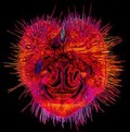
- Digital Images
- Online
Mouse nose, transverse section
David Linstead
- Digital Images
- Online
Transverse section through mouse soleus muscle
James N. Sleigh
- Digital Images
- Online
Blood vessel in a melanoma, SEM
S. Gschmeissner, K. Hodivala-Dilke & M. Stone
- Digital Images
- Online
Movie: 3D adult mouse skull
Kevin Mackenzie, University of Aberdeen
- Digital Images
- Online
Murine pancreas, SPIM
Jürgen Mayer, Centre de Regulació Genòmica & Universitat Pompeu Fabra
- Digital Images
- Online
Brown Adipose tissue, murine, THG
Daniela Malide, NIH, Bethesda, USA
- Digital Images
- Online
Mouse blastocyst 3.5 days after fertilisation
Lyndsey Butterworth, Newcastle University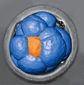
- Digital Images
- Online
Early mouse embryo preimplantation, LM
Rajeev Samarage, Melanie White, Andreas Fouras and Nicolas Plachta, Monash University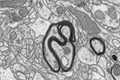
- Digital Images
- Online
Myelinated nerves in a mouse brain, TEM
Mikaela Laine, University of Helsinki
- Digital Images
- Online
Cartilage, trabecular bone and bone marrow in a mouse femur
Kevin Mackenzie, University of Aberdeen
- Digital Images
- Online
Mouse colon
Paul Appleton, University of Dundee
- Digital Images
- Online
Myelinated nerves in a mouse brain, TEM
Mikaela Laine, University of Helsinki
- Digital Images
- Online
Neuromuscular junctions and blood vessels
James N. Sleigh
- Digital Images
- Online
Mouse embryo
Macroscopic Solutions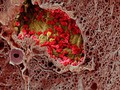
- Digital Images
- Online
Blood vessel in a melanoma, SEM
S. Gschmeissner, K. Hodivala-Dilke & M. Stone