Myelinated nerves in a mouse brain, TEM

  • Mikaela Laine, University of Helsinki
  • Digital Images
  • Online

Available online

view Myelinated nerves in a mouse brain, TEM

Attribution 4.0 International (CC BY 4.0)

You can use this work for any purpose, including commercial uses, without restriction under copyright law. You should also provide attribution to the original work, source and licence. Read more about this licence.

Credit

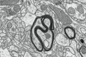
Myelinated nerves in a mouse brain, TEM. Mikaela Laine, University of Helsinki. Attribution 4.0 International (CC BY 4.0). Source: Wellcome Collection.

Selected images from this work

View 1 image

About this work

Description

Transmission electron micrograph of tissue from the medial prefrontal cortex of a mouse brain. This region of the brain is important for social behaviour, emotions and psychiatric disorders such as depression. In the centre of the image are two nerve fibres (axons) each surrounded by myelin (thick black line). Axons are long projections which extend out from nerve cells (neurones) and carry electrical impulses or signals to other cells. Myelin forms a protective sheath or insulating layer around some nerve fibres. It allows electrical impulses to travel quickly along the nerve cells. If it is damaged these impulses slow down and can cause diseases such as multiple sclerosis. Width of image is approximately 4 micrometres.

Permanent link