197 results filtered with: Digital Images

- Digital Images
- Online
Ca/cell cycle changes,Drosophila devt. movie
Huw Parry & Michael Whitaker
- Digital Images
- Online
Calcium/cell cycle changes, Drosophila development movie
Huw Parry & Michael Whitaker
- Digital Images
- Online
Calcium/cell cycle changes, Drosophila development movie
Huw Parry & Michael Whitaker
- Digital Images
- Online
Calcium/cell cycle changes, Drosophila development movie
Huw Parry & Michael Whitaker
- Digital Images
- Online
Calcium/cell cycle changes, Drosophila development movie
Huw Parry & Michael Whitaker
- Digital Images
- Online
Calcium/cell cycle changes, Drosophila development movie
Huw Parry & Michael Whitaker
- Digital Images
- Online
Calcium/cell cycle changes, Drosophila development movie
Huw Parry & Michael Whitaker
- Digital Images
- Online
Calcium/cell cycle changes, Drosophila development movie
Huw Parry & Michael Whitaker
- Digital Images
- Online
Calcium/cell cycle changes, Drosophila development movie
Huw Parry & Michael Whitaker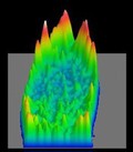
- Digital Images
- Online
Calcium/cell cycle changes, Drosophila development movie
Huw Parry & Michael Whitaker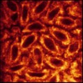
- Digital Images
- Online
Drosophila development, endoplasmic reticulum movie
Huw Parry & Michael Whitaker
- Digital Images
- Online
Drosophila development, nuclei/endoplasmic reticulum movie
H Parry, J Tweedie & M Whitaker
- Digital Images
- Online
Drosophila development endo.reticulum movie
Huw Parry & Michael Whitaker
- Digital Images
- Online
Drosophila development, nuclei/endoplasmic reticulum movie
H Parry, J Tweedie & M Whitaker
- Digital Images
- Online
Healthy human adult brain viewed from the side, tractography
Dr Flavio Dell'Acqua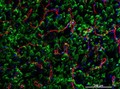
- Digital Images
- Online
Cellular architecture of normal human skin imaged by whole mount tissue microscopy. Human skin has a rich network of white blood cells (specifically dendritic cells, T cells and macrophages) which form sheaths around blood vessels. This image was taken directly beneath the junction that joins the dermal and epidermal layers of the skin (dermo-epidermal junction). At this level, the capillary network (stained for CD31; red) is visualised against a lawn of autofluorescent dermal papillae (finger-like projections of the dermis; green) scattered with dendritic cells (stained for CD11c; green) and macrophages (stained for LYVE-1; blue). This normal cellular architecture is grossly disrupted in diseased skin (see related images). Scale bar (white) represents 200 micrometres.
Dr. Xiao-nong Wang, Human Dendritic Cell Laboratory, Newcastle University
- Digital Images
- Online
Drosophila development, calcium/histones movie
Huw Parry & Michael Whitaker
- Digital Images
- Online
Drosophila adipose tissue
Christin Bauer
- Digital Images
- Online
GFP fluorescence studies, transfected cells
Biosciences Imaging Gp, Soton
- Digital Images
- Online
GFP fluorescence studies, transfected cells
Biosciences Imaging Gp, Soton
- Digital Images
- Online
Cell division
Kuan-Chung Su, London Research Institute, Cancer Research UK
- Digital Images
- Online
GFP fluorescence studies, untransfected cell
Biosciences Imaging Gp, Soton
- Digital Images
- Online
GFP fluorescence studies, untransfected cell
Biosciences Imaging Gp, Soton
- Digital Images
- Online
Staurosporine disrupts neutrophil membranes
A. Walker, L. Sharp & J. Pryde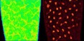
- Digital Images
- Online
Drosophila development, calcium & histones
Huw Parry & Michael Whitaker