654 results
- Pictures
Stomach cancer in a 70-year old man with a defect in the stomach wall where a gastroenterostomy was perfomed to remove an ulcer: showing fungating carcinoma tumour growing on the left. Pen and ink drawing by Barbara E. Nicholson, 1953.
Nicholson, BarbaraDate: 1953Reference: 34626iPart of: Barbara Nicholson medical illustration collection.- Archives and manuscripts
- Online
Burroughs Wellcome & Co, Private Letter Book 22
Date: September 1904 - March 1906Reference: WF/E/03/22Part of: Wellcome Foundation Ltd- Pictures
Operation to remove hernia from a female patient, showing the incision (above the pubis) with the hernia sac being removed, revealing the iliac vien and the commencement of repair, to unite two surrounding ligaments with surgical needle. Pencil drawing by Barbara E. Nicholson, 1947.
Nicholson, BarbaraDate: 1947Reference: 32189iPart of: Barbara Nicholson medical illustration collection.- Pictures
Chronic gastric ulcer, infiltrated with tumours in 68-year old man with fatal bronchopneumonia. Pencil drawing by Barbara E. Nicholson, 1947.
Nicholson, BarbaraDate: 1947Reference: 31874iPart of: Barbara Nicholson medical illustration collection.- Archives and manuscripts
- Online
Henry Wellcome Letter Book 3 ['Letter Book 3']
Date: Mar 1890 - Nov 1896Reference: WF/E/01/01/03Part of: Wellcome Foundation Ltd- Archives and manuscripts
- Online
Henry Wellcome Letter Book 4 ['Letter Book HSW Personal 2']
Date: Nov 1896 - Jan 1899Reference: WF/E/01/01/04Part of: Wellcome Foundation Ltd- Archives and manuscripts
- Online
Henry Wellcome Letter Book 6
Date: Aug 1901 - Jul 1903Reference: WF/E/01/01/06Part of: Wellcome Foundation Ltd- Pictures
Distended, congenitally defective, kidney malformed in the shape of a horse-shoe in a 55-year old man with thrombosis. Pencil drawing by Barbara E. Nicholson, 1947.
Nicholson, BarbaraDate: 1947Reference: 32101iPart of: Barbara Nicholson medical illustration collection.- Pictures
Intestinal cancer and mesenteric thrombosis in a 68-year old man with infarcted gut and gangrene: diagram demonstrating conduction of blood in relation to (a) venous obstruction and (b) tumorous occlusion. Pen and ink sketch by Barbara E. Nicholson, 1957.
Nicholson, BarbaraDate: 1957Reference: 35827iPart of: Barbara Nicholson medical illustration collection.- Pictures
Herpes zoster in a 48-year old man: sketch of buttocks and legs, anterior view. Pen and ink drawing by Barbara E. Nicholson, 1950.
Nicholson, BarbaraDate: 1950Reference: 33899iPart of: Barbara Nicholson medical illustration collection.
- Pictures
- Online
Spores of Bacillus pestis which caused the plague and its vector the human flea (Pulex irritans). Coloured drawing by A.J.E. Terzi.
Terzi, A. J. E. (Amedeo John Engel), 1872-1956.Reference: 41552i- Pictures
Fracture of nasal bones in a girl: lateral and medial aspects of face. Pencil drawing by Barbara E. Nicholson, 1955.
Nicholson, BarbaraDate: 1955Reference: 35403iPart of: Barbara Nicholson medical illustration collection.- Pictures
Operation to remove hernia from a female patient, showing the incision (above the pubis) with the hernia sac attached to the canal just external to the cavity in Gimbernat's ligament. Pencil drawing by Barbara E. Nicholson, 1947.
Nicholson, BarbaraDate: 1947Reference: 32188iPart of: Barbara Nicholson medical illustration collection.- Pictures
Dissecting aortic aneurysm in a 46-year old man with dyspnoea and congestive heart failure. Watercolour by Barbara E. Nicholson, 1952.
Nicholson, BarbaraDate: 1952Reference: 34437iPart of: Barbara Nicholson medical illustration collection.
- Pictures
- Online
Hemispherectomy in an adult patient: four figures. Carbon dust drawing by A.J. Arnott, 1960.
Arnott, Audrey J.Date: May 6th 1960Reference: 780079i- Pictures
Splenic abscesses in a male patient with a haematoma in right upper abdomen. Pen and ink preparatory sketch by Barbara E. Nicholson, 1953.
Nicholson, BarbaraDate: 1953Reference: 34850iPart of: Barbara Nicholson medical illustration collection.
- Pictures
- Online
Cerebrospinal rhinorrhoea. Carbon dust drawing by A.J. Arnott.
Arnott, Audrey J.Date: [20th century]Reference: 780078i- Archives and manuscripts
Frederick John Warburton Thompson: papers
Thompson, Frederick John Warburton (d. c.1951)Date: 1900sReference: MS.9294
- Pictures
A human anatomical figure. Drawing, Nepalese, ca. 1800 (?).
Date: 1800Reference: 574912i
- Pictures
- Online
The muscles of the left leg, seen from the front, and the bones and muscles of the right leg seen in right profile, and between them, a patella. Drawing by Michelangelo Buonarroti, ca. 1515-1520.
Michelangelo Buonarroti, 1475-1564.Date: [1515?-1520?]Reference: 26058i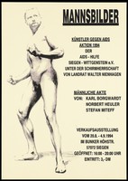
- Pictures
- Online
A naked man standing as if preparing to pull back; a drawing from an exhibition and auction in Siegen entitled: "Artists against AIDS" organized by AIDS-Hilfe Siegen Wittgenstein. Lithograph, 1994.
Date: 1994Reference: 673449i- Pictures
Fatal cancer tumour in right side of neck, in twelve year old girl. Pencil sketch by Barbara E. Nicholson, 1949.
Nicholson, BarbaraDate: 1949Reference: 33251iPart of: Barbara Nicholson medical illustration collection.- Pictures
Untitled.
Shrigley, DavidDate: [2020]Reference: 3272403iPart of: Wellcome Collection On Happiness Commision.- Pictures
Untitled.
Shrigley, DavidDate: [2020]Reference: 3272402iPart of: Wellcome Collection On Happiness Commision.- Pictures
Untitled.
Shrigley, DavidDate: [2020]Reference: 3272405iPart of: Wellcome Collection On Happiness Commision.