74 results
- Audio
A history of the brain. Part 6, The beast within.
Date: 2011
- Books
- Online
Demonstration of dog Louisa who reacts to acoustic stimuli after extirpation of both temporal lobes / by Walter B. Swift, A.B., S.B., M.D., assistant in neurology, Tufts Medical School; assistant to physcians for nervous diseases, Boston City Hospital.
Swift, Walter Babcock, 1868-Date: 1912
- Pictures
- Online
Head of "a backward boy" divided into four cerebral lobes: profile. Ink drawing with watercolour, c. 1900.
Date: c. 1900Reference: 28384i- Books
How inferior temporal cortex became a visual area / Charles G. Gross.
Gross, Charles G.Date: 1994
- Pictures
- Online
Two sections of the brain, divided into different lobes and faculties, according to Hollander's system of phrenology. Pen drawing, c. 1902.
Date: 1902Reference: 27965i
- Pictures
- Online
Head showing the 'convolutions' (lobes) of the brain. Pen drawing after R. W. Reid.
Reference: 28366i- Pictures
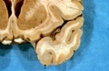
Cerebral haemorrhage and cystic kidneys in a 47-year old man: (a) brain section, showing clot in relation to temporal lobes, (b) right kidney section, revealing honeycombed architectural-like structures and (c) drawing of left kidney specimen, covered in carbuncular swellings. Watercolour by Barbara E. Nicholson, 1958.
Nicholson, BarbaraDate: 1958Reference: 36028iPart of: Barbara Nicholson medical illustration collection.
- Digital Images
- Online
Alzheimer’s Disease is a degenerative disorder of the brain which starts in middle or late life. It mainly affects the frontal and temporal lobes of the brain, where the cortex becomes atrophied. Plaques containing an amyloid-like protein have been found within the cortex on examination.
Susan Lockhart
- Pictures
- Online
Three diagrams of the organisation of the lobes of the brain for a phrenological textbook. Pen drawing, c. 1902.
Date: 1902Reference: 27967i- Pictures
A skull with a high parietal bone; another indicating diminished frontal and enlarged occipital lobes. 2 photomechanical reproductions, c. 1902.
Date: 1902Reference: 28072i
- Digital Images
- Online
Brain: tuberculous meningitis
- Digital Images
- Online
Nocardiosis: intracranial abscess
- Archives and manuscripts
Kreiman, Fried and Koch
Date: 2002Reference: PP/CRI/N/11/2/5Part of: Francis Crick (1916-2004): archives
- Digital Images
- Online
White matter fibres of the uncinate fasciculus
Christopher Whelan, Royal College of Surgeons in Ireland
- Digital Images
- Online
White matter fibres of the uncinate fasciculus
Christopher Whelan, Royal College of Surgeons in Ireland
- Pictures
- Online
The human brain, divided according to Bernard Hollander's system of phrenology. Process print with pen and ink, c. 1902.
Date: [approximately 1902]Reference: 27959i- Archives and manuscripts
- Online
'Hard copy of slides'
Date: 2000-2001Reference: PP/CRI/L/1/5/13Part of: Francis Crick (1916-2004): archives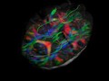
- Digital Images
- Online
White matter fibres of the uncinate fasciculus
Christopher Whelan, Royal College of Surgeons in Ireland
- Digital Images
- Online
White matter fibres of the uncinate fasciculus
Christopher Whelan, Royal College of Surgeons in Ireland
- Digital Images
- Online
White matter fibres of the uncinate fasciculus
Christopher Whelan, Royal College of Surgeons in Ireland
- Digital Images
- Online
White matter fibres of the uncinate fasciculus
Christopher Whelan, Royal College of Surgeons in Ireland
- Digital Images
- Online
Brain: cerebral toxoplasmosis

- Digital Images
- Online
White matter fibres of the uncinate fasciculus
Christopher Whelan, Royal College of Surgeons in Ireland
- Digital Images
- Online
White matter fibres of the uncinate fasciculus
Christopher Whelan, Royal College of Surgeons in Ireland- Archives and manuscripts
Miscellaneous accumulated papers on psychiatry
Date: c.1955-1958Reference: PP/ROS/C/3/4Part of: The Archive of Ismond Rosen (1924-1996)