75 results

- Archives and manuscripts
- Online
Ultra-violet microscope image referenced as "Newt metaphase U.V. (copy of original)"
Wilkins, Maurice Hugh Frederick, 1916-2004Date: February 1951Reference: KDBP/1/1/0262Part of: King's College London Department of Biophysics
- Digital Images
- Online
Human chromosomes in metaphase. The chromosomes are all aligned and at this stage they are attached to the spindle (not visible in this image).
Matthew Daniels
- Digital Images
- Online
Human cells showing the stages of cell division starting with interphase at the top. Progressing down, the stages shown are: prophase, metaphase (chromosomes all attached and aligned), anaphase (chromosome separation)and telophase (formation of midbody and cells begin to flatten).
Matthew Daniels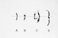
- Digital Images
- Online
Chromosome condensation prophase to metaphas
Wessex Reg. Genetics Centre
- Digital Images
- Online
Human chromosomes in metaphase. The chromatin is stained red and the "glue" that holds the two chromatids together is highlighted in yellow. This glue is a proteinaceous complex called cohesin. Once all the chromosomes are attached to the spindle, the cohesin complex breaks down, allowing the two chromatids to separate and move to opposite poles of the cell.
Matthew Daniels
- Ephemera
- Online
Biovation : Biovation fluorescent chromosome paints and probes from Scotlab / Ford Kennedy.
Kennedy, Ford.Date: 1995
- Digital Images
- Online
Human cells showing the stages of cell division starting with interphase second from the left on the top. Progressing anticlockwise the stages shown are: early prophase (centrosome not yet separated), late prophase (centrosome separated and DNA condensation), prometaphase (incomplete chromosome attachment), metaphase (chromosomes all attached and aligned), anaphase (chromosome separation), telophase (formation of midbody and cells begin to flatten), early cytokinesis (chromosomes decondensed and nuclear envelope reformed) and late cytokinesis (cells move apart).
Matthew Daniels
- Ephemera
- Online
Fluorescent chromosome paints / Scotlab.
Scotlab (Firm)Date: [1995]
- Digital Images
- Online
Cosmid DNA probe for chromosome 9, human
Dr Rosemary Ekong, UCL
- Digital Images
- Online
Catharanthus roseus (L.)G.Don Apocynaceae. Madagascar Periwinkle Distribution: Madagascar. It is the source of vincristine and vinblastine, which impair cell multiplication by interfering with microtubule assembly, causing metaphase arrest and are effective medications for leukaemias, lymphomas and some solid tumours. The mortality from childhood leukaemia fell from 100% to 30% once it was introduced - not a drug that could ethically be tested by double-blind trials. These chemicals were initially discovered by investigators in 1958 who were looking for cures for diabetes so tested this plant which was being used in the West Indies to reduce blood sugar levels. There are 70 different alkaloids present in this plant, and some - catharanthine, leurosine sulphate, lochnerine, tetrahydroalstonine, vindoline and vindolinine - lower blood sugar levels. However, the toxicity of this plant is such that this is not a plant to try at home for diabetic management. The vincristine content of the plant is 0.0003%, so two kilograms of leaf are required to produce sufficient vincristine for a single course of treatment for a child (6gm). Fortunately it is a vigorous weed and easy to grow in the tropics. Artificial synthesis has now been achieved. Photographed in the Medicinal Garden of the Royal College of Physicians, London.
Dr Henry Oakeley
- Digital Images
- Online
DNA probe for Y chromosome, meta/interphase
Dr Rosemary Ekong, UCL
- Digital Images
- Online
Human chromosomes at three stages of mitosis
Laura Trinkle-Mulcahy
- Digital Images
- Online
Meiosis in an oocyte
M. Herbert, A. McDougall & H. Homer
- Digital Images
- Online
Foetal cells in amniotic fluid, + colchicine
Wessex Reg. Genetics Centre
- Digital Images
- Online
Location of cyclin in 2-cell human embryo
Dr Mark Carrington,Camb.Univ.
- Digital Images
- Online
Location of cyclin in 2-cell human embryo
Dr Mark Carrington,Camb.Univ.
- Digital Images
- Online
Location of cyclin in 2-cell human embryo
Dr Mark Carrington,Camb.Univ.
- Digital Images
- Online
Foetal cells from amniotic fluid, colchicine
Wessex Reg. Genetics Centre
- Digital Images
- Online
Location of cyclin in maturing human egg
Dr Mark Carrington,Camb.Univ.
- Digital Images
- Online
Location of cyclin in maturing human egg
Dr Mark Carrington,Camb.Univ.
- Digital Images
- Online
Location of cyclin in maturing human egg
Dr Mark Carrington,Camb.Univ.
- Digital Images
- Online
Translocation shown up by cosmid probes
Wessex Reg. Genetics Centre
- Digital Images
- Online
Human HeLa cancer cells, stages of mitosis
William J Moore/Univ. Dundee
- Digital Images
- Online
Human chromosomes during cell division
Matthew Daniels
- Digital Images
- Online
DNA probe for human/hamster hybrid DNA
Dr Rosemary Ekong, UCL