1,341 results
- Archives and manuscripts
- Online
"2nd Layer Line Straight"
Date: c.1957Reference: FRKN 2/28Part of: The Papers of Rosalind Franklin
- Books
- Online
The nature of the intra-ocular fluids / by W. Stewart Duke-Elder ; being the Sir Francis Laking Prize, 1926-1927, from the Biochemical Department, St. George's Hospital London.
Duke-Elder, Stewart, Sir, 1898-1978.Date: 1827- Archives and manuscripts
Lay committee applications
Date: 2002Reference: SA/FIH/A/1/2/2Part of: Foundation for Integrated Health- Archives and manuscripts
- Online
Adhoc committee on training of Lay Workers
Date: Nov 1961-Oct 1964Reference: SA/FPA/A19/39Part of: Family Planning Association
- Digital Images
- Online
Bowman's layer and inflammatory cells
Rob Young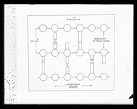
- Archives and manuscripts
- Online
Drawing of the layer structure model of collagen referenced as "Layer structure model. Collagen"
Franklin, Rosalind, 1920-1958Date: March 1952Reference: KDBP/1/1/0607Part of: King's College London Department of Biophysics- Archives and manuscripts
- Online
"1st Layer Line"
Date: Jul 1958Reference: FRKN 2/11Part of: The Papers of Rosalind Franklin- Archives and manuscripts
- Online
"7th Layer Line"
Date: 1957Reference: FRKN 2/27Part of: The Papers of Rosalind Franklin- Archives and manuscripts
- Online
"5th Layer Line"
Date: Feb 1956Reference: FRKN 2/25Part of: The Papers of Rosalind Franklin- Archives and manuscripts
"4th Layer Line"
Date: Mar 1956Reference: FRKN 2/10Part of: The Papers of Rosalind Franklin- Archives and manuscripts
Layer V 1992-1993
Date: 1977-1993Reference: PP/CRI/L/1/4/5Part of: Francis Crick (1916-2004): archives- Archives and manuscripts
- Online
"6th Layer Line"
Date: Jan 1957Reference: FRKN 2/16Part of: The Papers of Rosalind Franklin- Archives and manuscripts
- Online
"2nd Layer Line"
Date: 1953-1968Reference: FRKN 2/8Part of: The Papers of Rosalind Franklin- Archives and manuscripts
- Online
"9th Layer Line"
Date: c.1954-1957Reference: FRKN 2/6Part of: The Papers of Rosalind Franklin
- Books
- Online
The butterfly vivarium, or, Insect home: being an account of a new method of observing the curious metamorphoses of some of the most beautiful of our native insects. Comprising also a popular description of the habits and instincts of many of the insects of the various classes referred to; with suggestions for the successful study of entomology by means of an insect vivarium / by H. Noel Humphreys ... Illustrated with coloured engravings.
Humphreys, Henry Noel, 1810-1879.Date: 1858- Archives and manuscripts
Lay committee correspondence and applications
Date: 2005-2007Reference: SA/FIH/A/1/2/3Part of: Foundation for Integrated Health
- Pictures
- Online
Anatomy: écorché human back, showing cutaneous nerves and the first layer of muscle. Photograph, ca. 1900.
Waterston, D. (David)Date: 1900Reference: 575097i- Archives and manuscripts
Lay involvement: Applications for position of chair
Date: 2002-2006Reference: SA/FIH/A/1/2/1Part of: Foundation for Integrated Health
- Digital Images
- Online
Tortoise: shell burn - horny layer destroyed
Royal Veterinary College- Books
Dünnschichtchromatographie in der Aminosäure- und Peptid-Chemie / von György Pataki.
Pataki, György.Date: 1966- Archives and manuscripts
- Online
"3rd Layer Line Photometer Curve"
Date: Feb 1956Reference: FRKN 2/17Part of: The Papers of Rosalind Franklin
- Digital Images
- Online
Layer of epithelial cells - coloured
University of Edinburgh- Archives and manuscripts
- Online
"3rd Layer Line Helical Projection"
Date: 1957Reference: FRKN 2/18Part of: The Papers of Rosalind Franklin
- Archives and manuscripts
- Online
Template referenced as "2nd layer plane"
Wilkins, Maurice Hugh Frederick, 1916-2004Date: November 1953Reference: KDBP/1/1/1233Part of: King's College London Department of Biophysics
- Digital Images
- Online
Brain with a thick layer of pus on its surface
Godart, Thomas