53 results filtered with: Model organism

- Digital Images
- Online
Transgenic Drosophila pupa expressing GFP
Derric Nimmo & Paul Eggleston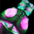
- Digital Images
- Online
Dorsal view of zebrafish brain (4 day-post fertilization)
Anya Suppermpool, Rihel lab/ Wilson lab, University College London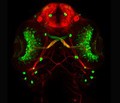
- Digital Images
- Online
Eye development, zebrafish
Kate Turner, Dr Steve Wilson
- Digital Images
- Online
Zebrafish posterior lateral line development
Leo Valdivia, Dr Steve Wilson
- Digital Images
- Online
Transverse section of brain and eyes, Zebrafish model
Leo Valdivia, Dr Steve Wilson
- Digital Images
- Online
Retina development, composition
Leo Valdivia, Dr Steve Wilson
- Digital Images
- Online
Liver of a DEN (Diethylnitrosamine)-treated rat. DEN is a toxic chemical which quickly induces liver cirrhosis followed by HCC (Hepatocellular carcinoma, a primary liver cancer). Cirrhosis is an end result of fibrosis, the scarring of liver tissue. Fibrosis is caused by the overproduction of collagen, a component of the connective tissue forming the liver. To grade the amount of cirrhosis present in a liver sample, collagen is made visible using the dye sirius red. Under polarized light, collagen is observed as the golden to red color as shown in this image.
Tabea Hohensee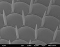
- Digital Images
- Online
Drosophila eye
Anne Weston, Francis Crick Institute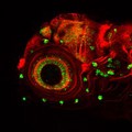
- Digital Images
- Online
Zebrafish sensory neuromasts
Kate Turner, Dr Steve Wilson
- Digital Images
- Online
Zebrafish embryos with green fluorescent myotomes
S. Roy & S Higashijima
- Digital Images
- Online
Transgenic Drosophila pupa expressing GFP in its eyes
Derric Nimmo & Paul Eggleston
- Digital Images
- Online
Transgenic Drosophila pupa expressing GFP
Derric Nimmo & Paul Eggleston
- Digital Images
- Online
Transgenic Drosophila pupa expressing GFP
Derric Nimmo & Paul Eggleston
- Digital Images
- Online
Zebrafish embryos with green fluorescent notocords
S. Roy & F. Muller
- Digital Images
- Online
Drosophila proboscis
Anne Weston, Francis Crick Institute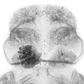
- Digital Images
- Online
Dorsal view of the forebrain of a wild-type zebrafish embryo
Ana Faro, Dr Steve Wilson
- Digital Images
- Online
GABAergic and Glutamatergic neurons in the zebrafish brain
Kate Turner, Dr Steve Wilson
- Digital Images
- Online
Zebrafish scales
Anne Weston, Francis Crick Institute
- Digital Images
- Online
Transgenic Drosophila expressing GFP in their eyes
Derric Nimmo & Paul Eggleston
- Digital Images
- Online
Dopaminergic neurons in the zebrafish forebrain. Confocal micrograph of a 4 day old transgenic zebrafish embryo viewed from a lateral aspect. Neurons in the olfactory bulb, telencepahlon, ventral diencephalon, pretectum and hypothalamus are labelled in green. Axonal tracts are shown in cyan and neuropil in magenta. In order to show the anatomy of the brain better the skin and eyes of the embryo have been removed post-fixation.
Kate Turner, Dr Steve Wilson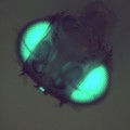
- Digital Images
- Online
Transgenic Drosophila expressing GFP in its eyes and ocelli
Derric Nimmo & Paul Eggleston
- Digital Images
- Online
Glycinergic neurons in a zebrafish embryo
Kate Turner, Dr Steve Wilson
- Digital Images
- Online
Arabidopsis thaliana plant cells containing chloroplasts, LM
Fernán Federici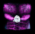
- Digital Images
- Online
Brain development, zebrafish
Ingrid Lekk, Dr Steve Wilson
- Digital Images
- Online
Zebrafish parapineal development
Ingrid Lekk, Dr Steve Wilson