34 results

- Digital Images
- Online
Interphase cells showing pentameric X
Wessex Reg. Genetics Centre- Archives and manuscripts
A diagram of "Interphase (DNA replication)" printed on cardboard.
Date: 20th centuryReference: SA/BIO/N/6Part of: The Biochemical Society
- Digital Images
- Online
Xenopus cancer kidney cells, interphase
Paul Andrews/Univ. Dundee
- Digital Images
- Online
Xenopus cancer kidney cells, interphase
Paul Andrews/Univ. Dundee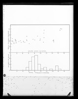
- Archives and manuscripts
- Online
Graph referenced as "Increase of Fuelgen staining during interphase"
Walker, Peter M. B.Date: November 1951Reference: KDBP/1/1/0562Part of: King's College London Department of Biophysics
- Digital Images
- Online
Nucleus in interphase. The large dark area is the nucleolus.
Matthew Daniels
- Archives and manuscripts
- Online
Graph referenced as "Increase of ultra-violet absorbing material during interphase"
Walker, Peter M. B.Date: November 1951Reference: KDBP/1/1/0558Part of: King's College London Department of Biophysics
- Archives and manuscripts
- Online
Graph referenced as "Protein/DNA. During interphase. Mouse heart t.e. cells"
Richards, E. G. (Edward Graham)Date: November 1957Reference: KDBP/1/1/2447Part of: King's College London Department of Biophysics
- Books
- Online
The mitotic cycle : the cytoplasm and nucleus during interphase and mitosis / by Arthur Hughes.
Hughes, Arthur Frederick William, 1908-Date: 1952
- Digital Images
- Online
Two human cells in interphase. The microtubules are stained in green and the DNA in red.
Matthew Daniels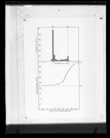
- Archives and manuscripts
- Online
Graph recording levels of DNA during interphase in a mouse sample referenced as "DNA synthesis mouse t.e."
Richards, E. G. (Edward Graham)Date: August 1955Reference: KDBP/1/1/1705Part of: King's College London Department of Biophysics- Archives and manuscripts
- Online
'Gene order within 9q32-34 using interphase analysis by fluorescence in situ hydridisation' with Margaret A Leversha et al.
Date: 1992Reference: UGC 188/4/1/8/3Part of: Papers of Malcolm Andrew Ferguson-Smith, geneticist, Professor of Medical Genetics, University of Glasgow, Scotland
- Digital Images
- Online
Human cell in interphase showing the tubulin component of the cytoskeleton in green, the DNA in blue and the kinetochores in pink.
Matthew Daniels
- Digital Images
- Online
Human cell in interphase showing the tubulin component of the cytoskeleton in green and the DNA in red. The centrosome, to which the microtubules attach, can be seen to the right of the nucleus.
Matthew Daniels
- Digital Images
- Online
Human cells showing the stages of cell division starting with interphase at the top. Progressing down, the stages shown are: prophase, metaphase (chromosomes all attached and aligned), anaphase (chromosome separation)and telophase (formation of midbody and cells begin to flatten).
Matthew Daniels
- Ephemera
- Online
Biovation : Biovation fluorescent chromosome paints and probes from Scotlab / Ford Kennedy.
Kennedy, Ford.Date: 1995
- Digital Images
- Online
Human cells showing the stages of cell division starting with interphase second from the left on the top. Progressing anticlockwise the stages shown are: early prophase (centrosome not yet separated), late prophase (centrosome separated and DNA condensation), prometaphase (incomplete chromosome attachment), metaphase (chromosomes all attached and aligned), anaphase (chromosome separation), telophase (formation of midbody and cells begin to flatten), early cytokinesis (chromosomes decondensed and nuclear envelope reformed) and late cytokinesis (cells move apart).
Matthew Daniels
- Ephemera
- Online
Fluorescent chromosome paints / Scotlab.
Scotlab (Firm)Date: [1995]
- Digital Images
- Online
DNA probe for Y chromosome, meta/interphase
Dr Rosemary Ekong, UCL- Film
- Online
Portio-carcinoma in vitro : type hela : homo sapiens cytomorphology.
Date: 1961
- Digital Images
- Online
Human HeLa cancer cells, stages of mitosis
William J Moore/Univ. Dundee
- Digital Images
- Online
Human chromosomes during cell division
Matthew Daniels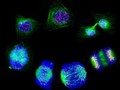
- Digital Images
- Online
Human cells showing the stages of cell division
Matthew Daniels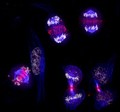
- Digital Images
- Online
Human HeLa cancer cells, mitosis
Paul Andrews/Univ. Dundee
- Digital Images
- Online
Drosophila development, effect of heparin movie
Huw Parry & Michael Whitaker