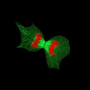
High resolution confocal micrograph of a dividing HeLa cell. Condensed chromosomes (red) have aligned and attached to cytoskeleton microtubules which form the spindle (green). During anaphase (one of the stages of nuclear division in mitosis), the spindle pulls the condensed chromosomes to opposite poles of the cells. During the next stage, telophase, new nuclear membranes form around the two groups of separated chromosomes. This is followed by cytokinesis, the process by which a cell divides its cytoplasm and physically separates the two daughter cells.
HeLa cells are an immortal human epithelial cell line derived from a cancerous tumour of the cervix (adenocarcinoma). It was established in 1951 from a biopsy taken from Henrietta Lacks, and was the first human cell line to survive and grow in the laboratory. Henrietta's cells were originally used in this way without permission from her or her family which raises issues about ethics and privacy. HeLa cells have been used extensively around the world in many different fields of research including cancer research, immunology and vaccine development. Width of image is 78 micrometres.