Stories
- Article
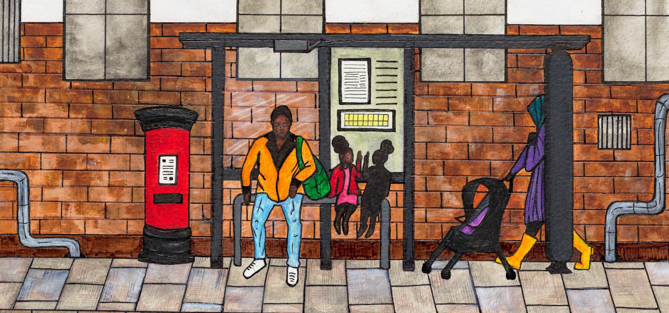
We need less ‘sickle cell warriors’ and more allies
Rejecting the epithet “warrior”, Cheryl Telfer describes the pervasive effect sickle cell disease has on her life, and calls for more people to donate blood to help sicklers.
- Article
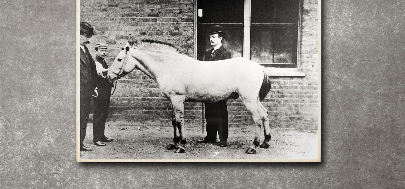
Jim, the horse of death
Horses’ blood was used to produce an antitoxin that saved thousands of children from dying from diphtheria, but contamination was a deadly problem. Find out how a horse called Jim was the catalyst for the beginnings of medical regulation.

- Article
The leukaemia diagnosis I didn’t see coming
Treatment for leukaemia kept journalist Hannah Partos in isolation, like the female prisoner whose image inspired her to write this piece.

- Article
Womb milk and the puzzle of the placenta
A human baby needs milk to survive – and this holds true even before it’s born. Joanna Wolfarth explores “womb milk”, as well as ancient and modern ideas about the placenta.


Catalogue

- Digital Images
- Online
Blood vessel with red and white blood cells
University of Edinburgh
- Digital Images
- Online
Blood vessel with red and white blood cells
University of Edinburgh
- Digital Images
- Online
Monocyte and two red blood cells
University of Edinburgh
- Digital Images
- Online
Blood clot with crenated red cells
Anne Weston, Francis Crick Institute
- Digital Images
- Online
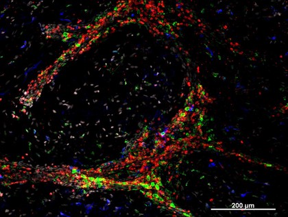
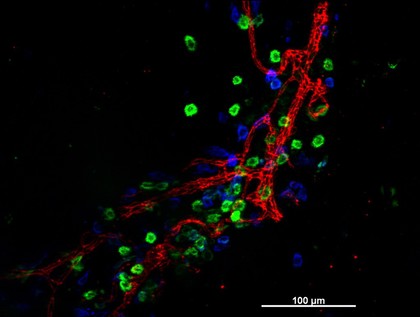
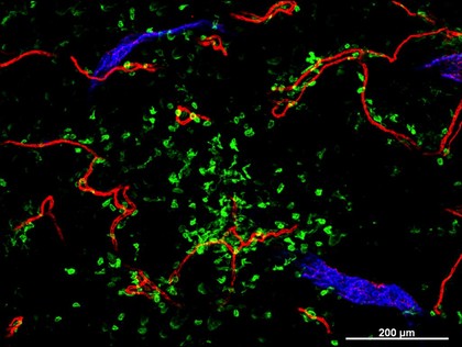
Cellular architecture of normal human skin imaged by whole mount tissue microscopy. Human skin has a rich network of white blood cells (specifically dendritic cells, T cells and macrophages) which form sheaths around blood vessels (string-like structures). A network of lymphatic vessels (ribbon-like structures) is also present. In this image, human skin lymphatic vessels (stained for LYVE-1; blue) and white blood cells comprised of dendritic cells (stained for CD11c; green) and T cells (stained for CD3; red) can be seen. Some macrophages also express the protein LYVE-1 similar to lymphatic vessel cells which can be appreciated as blue cells within and in between the sheaths of white blood cells. This normal cellular architecture is grossly disrupted in diseased skin (see related images). X10 magnification. Scale bar (white) represents 200 micrometres.
Dr. Xiao-nong Wang, Human Dendritic Cell Laboratory, Newcastle University