Stories

- Book extract
Dangers inside and out
Eimear McBride reflects on the deadly consequences of misogyny in the wake of the murder of Sarah Everard and argues why advising women to simply “stay indoors” is wrong.

- Article
Tripping for spiritualism and science
Getting high in the name of religion or creativity has been practised for centuries. Now it seems hallucinogenics could help treat mental illnesses too.
- Article
Stigma, schizophrenia and being transgender
When he was diagnosed with schizophrenia, Ashley McFord-Allister discovered that the medical world will not continue gender confirmation treatment while treating a mental health condition. Here he exposes the prejudice behind this attitude.
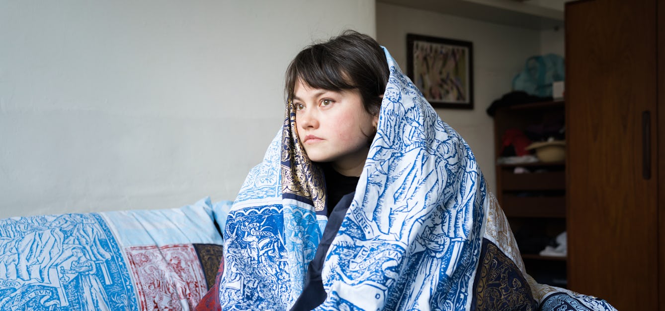
- Photo story
Beautiful bedding and how to die well
When you are unwell, your bed can be both a refuge and a prison. Discover how artist Poppy Nash created a bed-centred artwork inspired by her own chronic illness and depictions of ill health from history.


Catalogue
- Books
Standardization and quantitation of diagnostic staining in cytology / edited by Mathilde E. Boon and L.P. Kok.
Date: 1986- Books
The microtomist's formulary and guide / by Peter Gray.
Gray, Peter, 1908-1981.Date: 1954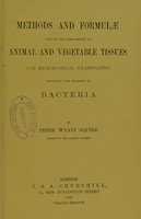
- Books
- Online
Methods and formulae used in the preparation of animal and vegetable tissues for microscopical examination : including the staining of bacteria / by Peter Wyatt Squire.
Date: 1892- Books
McClung's handbook of microscopial technique : for workers in animal and plant tissues / by thirty-five authors ; edited by Ruth McClung Jones.
Date: [1950], ©1950- Books
Physikochemische Grundlagen der histologischen Methodik / von K. Zeiger.
Zeiger, Karl, 1895-Date: 1938