Stories

- Book extract
The shape of thought
Santiago Ramón y Cajal’s description of the moment in 1887 when he saw a brain cell for the first time never fails to move neuroscientist Richard Wingate to tears. Here he captures that enduring sense of wonder.
- Article
The relationship between science and art
Often seen as opposites, science and art both depend on observation and synthesis.


Catalogue
- Archives and manuscripts
- Online
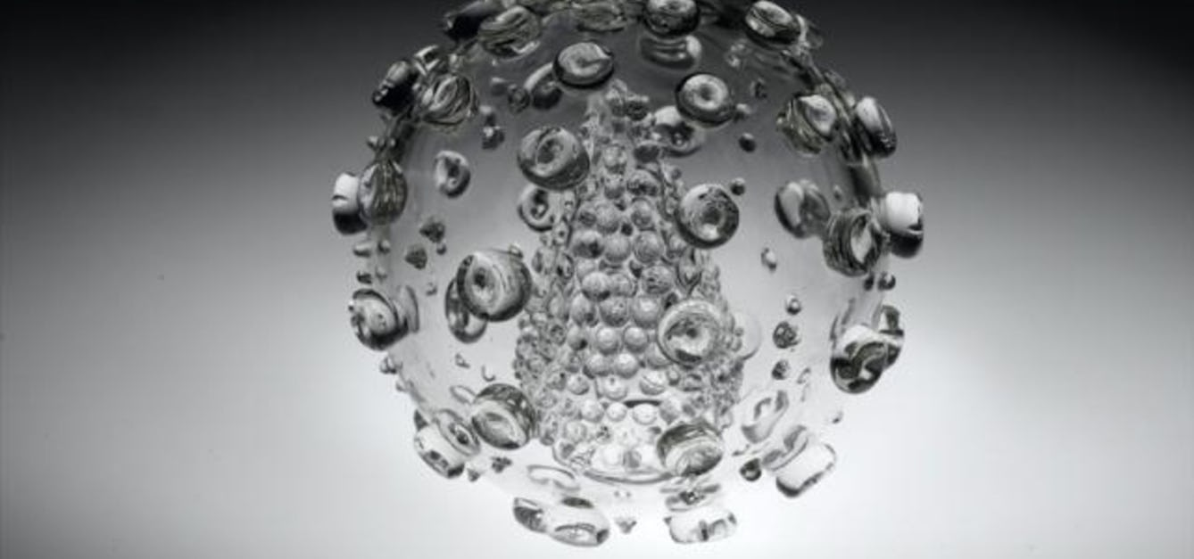
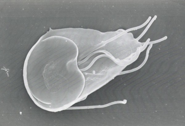
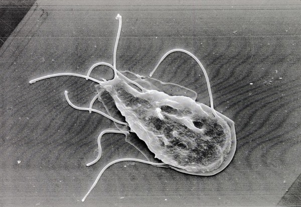
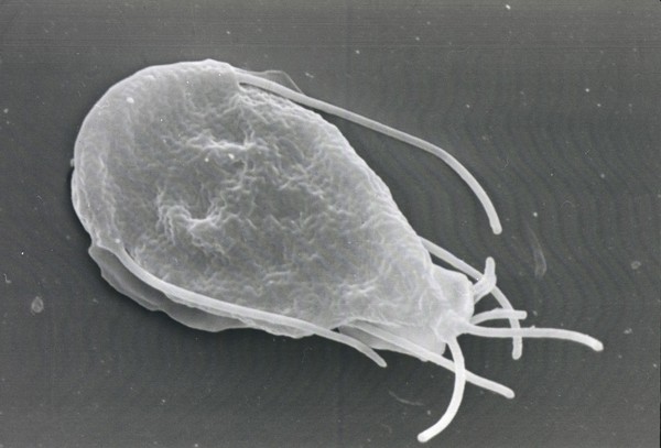
Electron Microscope Images
Date: 1950-1969Reference: SB/6/4/10Part of: Sydney Brenner Collection
- Archives and manuscripts
- Online
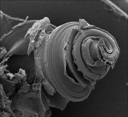
Electron microscope image referenced as "Belanger"
Pelc, Stephen RichardDate: February 1962Reference: KDBP/1/1/4239Part of: King's College London Department of Biophysics
- Archives and manuscripts
- Online
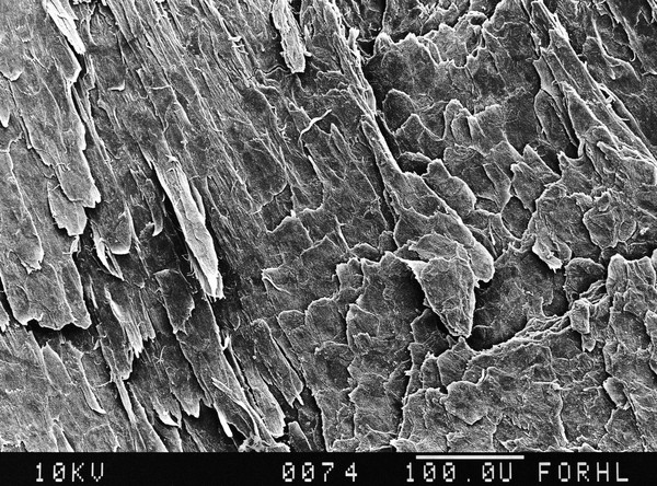
Electron microscope image referenced as "Bentonite trace"
McEwen, MargaretDate: July 1950Reference: KDBP/1/1/0191Part of: King's College London Department of Biophysics
- Archives and manuscripts
- Online
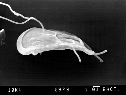
Electron microscope image referenced as "Rabbit sperm"
Bovey, R. (René)Date: July 1950Reference: KDBP/1/1/0193Part of: King's College London Department of Biophysics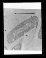
- Archives and manuscripts
- Online
Electron microscope image referenced as "Kellenberger"
Wilkins, Maurice Hugh Frederick, 1916-2004Date: August 1960Reference: KDBP/1/1/3492Part of: King's College London Department of Biophysics