


Catalogue
- Journals
Dapim refuiim : Folia medica.
Date: 1935-1969
- Digital Images
- Online
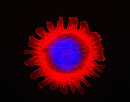
Cell flower formed from a 3T3 fibroblast cell. An unusually shaped cell found growing under normal conditions. The cell nucleus, containing the DNA, is stained in blue with DAPI. The cell body is stained for F-actin in red to reveal the flowere like shape.
Steve Winder
- Digital Images
- Online
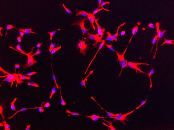
Human neural stem cells stained for nestin (red). Nestin is a type of intermediate filamant protein that is used as a marker of neural stem cells. The blue dots are the cell nuclei stained with DAPI. Neural stem cells can be made to develop into cells found in the central nervous system; neurons, astrocytes and oligodendrocytes.
Yirui Sun
- Digital Images
- Online
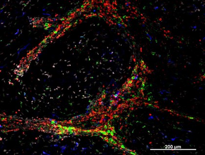
Cellular architecture of normal human skin imaged by whole mount tissue microscopy. Human skin has a rich network of white blood cells (specifically dendritic cells, T cells and macrophages) which form sheaths around blood vessels. In this image, T cells (stained for CD3; red) dendritic cells (stained for MHC class II; green) and macrophages (stained for LYVE-1; blue with some cells showing a tinge of green) can be seen. Cell nuclei have been stained with DAPI (grey). This normal cellular architecture is grossly disrupted in diseased skin (see related images). X10 magnification. Scale bar (white) represents 200 micrometres.
Dr. Xiao-nong Wang, Human Dendritic Cell Laboratory, Newcastle University
- Digital Images
- Online
Cellular architecture of normal human skin imaged by whole mount tissue microscopy. Human skin has a rich network of white blood cells (specifically dendritic cells, T cells and macrophages) which form sheaths around blood vessels. In this image, T cells (stained for CD3; red) dendritic cells (stained for MHC class II; green) and macrophages (stained for LYVE-1; blue with some cells showing a tinge of green) can be seen. Cell nuclei have been stained with DAPI (grey). This normal cellular architecture is grossly disrupted in diseased skin (see related images). X20 magnification. Scale bar (white) represents 100 micrometres.
Dr. Xiao-nong Wang, Human Dendritic Cell Laboratory, Newcastle University