302 results filtered with: Digital Images, Pictures

- Pictures
- Online
A group of men look up as they join arms in a ring representing an advertisement for the Positive Support Network for people living with HIV/AIDS by ACT PLWA and the AIDS Action Council. Colour lithograph.
Date: [between 1990 and 1999]Reference: 669749i
- Pictures
- Online
A Maori man holding a flaming torch; advertising the 11th Internernational AIDS Candlelight Memorial and Mobilization organized by Global AIDS Action Network on Sunday 22nd May, part of a project by the New Zealand AIDS Foundation. Colour lithograph by Stephen A'Court.
Date: [between 1990 and 1999]Reference: 669569i
- Pictures
- Online
The repeated words 'the right move' against a hazy image of a couple in bed representing an advertisement for safe sex by the Gay Men's Outreach Crew. Colour lithograph by Bradley Brechin.
Date: [between 1900 and 1999]Reference: 668336i
- Digital Images
- Online
Neural network
Arran Lewis
- Digital Images
- Online
Collagen network
David Gregory & Debbie Marshall
- Pictures
- Online
Silhouettes of people with hearts walking representing an advertisement for the AIDS Walk for Life in Edmonton Canada, Sunday Oct 4 1992. Colour lithograph after Keith Haring.
Haring, Keith.Date: Sunday Oct 4 1992Reference: 668423i
- Digital Images
- Online
Mitochondria network
Odra Noel
- Pictures
- Online
Silhouettes of figures with hearts walking representing an advertisement for the AIDS Walk for Life at Rundle Park Family Recreation Centre, Edmonton Canada, 3 Oct. 1993. Colour lithograph.
Date: Sunday Oct 3 1993Reference: 668116i
- Pictures
- Online
Components of the electromechanical telegraph network. Process print.
Reference: 473350i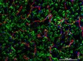
- Digital Images
- Online
Cellular architecture of normal human skin imaged by whole mount tissue microscopy. Human skin has a rich network of white blood cells (specifically dendritic cells, T cells and macrophages) which form sheaths around blood vessels. This image was taken directly beneath the junction that joins the dermal and epidermal layers of the skin (dermo-epidermal junction). At this level, the capillary network (stained for CD31; red) is visualised against a lawn of autofluorescent dermal papillae (finger-like projections of the dermis; green) scattered with dendritic cells (stained for CD11c; green) and macrophages (stained for LYVE-1; blue). This normal cellular architecture is grossly disrupted in diseased skin (see related images). Scale bar (white) represents 200 micrometres.
Dr. Xiao-nong Wang, Human Dendritic Cell Laboratory, Newcastle University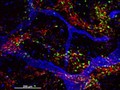
- Digital Images
- Online
Cellular architecture of normal human skin imaged by whole mount tissue microscopy. Human skin has a rich network of white blood cells (specifically dendritic cells, T cells and macrophages) which form sheaths around blood vessels (string-like structures). A network of lymphatic vessels (ribbon-like structures) is also present. In this image, human skin lymphatic vessels (stained for LYVE-1; blue) and white blood cells comprised of dendritic cells (stained for CD11c; green) and T cells (stained for CD3; red) can be seen. Some macrophages also express the protein LYVE-1 similar to lymphatic vessel cells which can be appreciated as blue cells within and in between the sheaths of white blood cells. This normal cellular architecture is grossly disrupted in diseased skin (see related images). X10 magnification. Scale bar (white) represents 200 micrometres.
Dr. Xiao-nong Wang, Human Dendritic Cell Laboratory, Newcastle University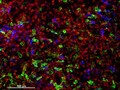
- Digital Images
- Online
Cellular architecture of human skin lymphoma imaged by whole mount tissue microscopy. Normal human skin has a rich network of white blood cells (specifically dendritic cells, T cells and macrophages) which form sheaths around blood vessels. In diseased skin, such as in skin lymphoma as seen here, this normal architecture becomes distorted. In this image, lots of T cells (stained for CD3; red), dendritic cells (stained for CD11c; green) and macrophages (stained for LYVE-1; blue) have infiltrated the skin. X20 magnification. Scale bar (white) represents 100 micrometres.
Dr. Xiao-nong Wang, Human Dendritic Cell Laboratory, Newcastle University
- Digital Images
- Online
Retinal capillary bed
Jean Wade and Linda Sharp
- Digital Images
- Online
Cellular architecture of normal human skin imaged by whole mount tissue microscopy. Human skin has a rich network of white blood cells (specifically dendritic cells, T cells and macrophages) which form sheaths around blood vessels. In this image, T cells (stained for CD3; red) dendritic cells (stained for MHC class II; green) and macrophages (stained for LYVE-1; blue with some cells showing a tinge of green) can be seen. Cell nuclei have been stained with DAPI (grey). This normal cellular architecture is grossly disrupted in diseased skin (see related images). X10 magnification. Scale bar (white) represents 200 micrometres.
Dr. Xiao-nong Wang, Human Dendritic Cell Laboratory, Newcastle University
- Digital Images
- Online
Cellular architecture of normal human skin imaged by whole mount tissue microscopy. Human skin has a rich network of white blood cells (specifically dendritic cells, T cells and macrophages) which form sheaths around blood vessels. In this image, T cells (stained for CD3; red) dendritic cells (stained for MHC class II; green) and macrophages (stained for LYVE-1; blue with some cells showing a tinge of green) can be seen. Cell nuclei have been stained with DAPI (grey). This normal cellular architecture is grossly disrupted in diseased skin (see related images). X20 magnification. Scale bar (white) represents 100 micrometres.
Dr. Xiao-nong Wang, Human Dendritic Cell Laboratory, Newcastle University
- Digital Images
- Online
Cellular architecture of normal human skin imaged by whole mount tissue microscopy. Human skin has a rich network of white blood cells (specifically dendritic cells, T cells and macrophages) which form sheaths around blood vessels. In this image, blood vessels (string-like structures stained for CD31; red), lymphatic vessels (ribbon-like structures stained for LYVE-1; blue) and dendritic cells (stained for CD11c; green) can be seen. Macrophages (stained for LYVE-1; blue) are also present. This normal cellular architecture is grossly disrupted in diseased skin (see related images). X10 magnification. Scale bar (white) represents 200 micrometres.
Dr. Xiao-nong Wang, Human Dendritic Cell Laboratory, Newcastle University
- Digital Images
- Online
Cellular architecture of normal human skin imaged by whole mount tissue microscopy. Human skin has a rich network of white blood cells (specifically dendritic cells, T cells and macrophages) which form sheaths around blood vessels. In this image, blood vessels (string-like structures stained for CD31; green), lymphatic vessels (ribbon-like structures stained for LYVE-1; blue) and T cells (stained for CD3; red) can be seen. T cells are only found around dermal blood vessels. Macrophages (stained for LYVE-1; blue) are also present. This normal cellular architecture is grossly disrupted in diseased skin (see related images). X10 magnification. Scale bar (white) represents 200 micrometres.
Dr. Xiao-nong Wang, Human Dendritic Cell Laboratory, Newcastle University
- Digital Images
- Online
Cellular architecture of normal human skin imaged by whole mount tissue microscopy. Human skin has a rich network of white blood cells (specifically dendritic cells, T cells and macrophages) which form sheaths around blood vessels. This image was taken greater than 150 micrometres beneath the junction that joins the dermal and epidermal layers of the skin (dermo-epidermal junction). At this level, dendritic cells (stained for CD11c; green) and macrophages (stained for LYVE-1; blue) form clusters around blood vessels (stained for CD31; red). This normal cellular architecture is grossly disrupted in diseased skin (see related images). Scale bar (white) represents 100 micrometres.
Dr. Xiao-nong Wang, Human Dendritic Cell Laboratory, Newcastle University
- Digital Images
- Online
Neurons in the brain
Dr Jonathan Clarke
- Digital Images
- Online
Human kidney cell, Gated-STED microscopy
Alison Dun, ESRIC (Edinburgh Super-Resolution Imaging Consortium)
- Digital Images
- Online
Cellular architecture of normal human skin imaged by whole mount tissue microscopy. Human skin has a rich network of white blood cells (specifically dendritic cells, T cells and macrophages) which form sheaths around blood vessels. This image was taken less than 20 micrometres beneath the junction that joins the dermal and epidermal layers of the skin (dermo-epidermal junction). At this level, dendritic cells (stained for CD11c; green) form clusters around and between blood capillary loops (stained for CD31; red). The blind-ended tips of initial lymphatic vessels are just visible (stained for LYVE-1; blue) at this level. This normal cellular architecture is grossly disrupted in diseased skin (see related images). Scale bar (white) represents 200 micrometres.
Dr. Xiao-nong Wang, Human Dendritic Cell Laboratory, Newcastle University
- Pictures
- Online
A dotted square bearing the words "The presence of absence"; advertising an exhibition of the Canadian AIDS Memorial Quilt in Edmonton at the Citadel Theatre, June 26 to July 5, 1995. Colour lithograph by Quality Colour Press and Cheryl Anne Lieberman Typographics.
Date: 1995Reference: 668184i
- Digital Images
- Online
Rat spinal cord, LM
Kevin Mackenzie, University of Aberdeen
- Digital Images
- Online
Osteocyte in cortical bone, SEM
Kevin Mackenzie, University of Aberdeen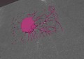
- Digital Images
- Online
Osteocyte in cortical bone, SEM
Kevin Mackenzie, University of Aberdeen