135 results filtered with: Digital Images
- Digital Images
- Online
The Microscopy and Zoology.

- Digital Images
- Online
The Microscopy and Zoology.
- Digital Images
- Online
The Microscopy and Zoology.

- Digital Images
- Online
Myxoid liposarcoma, microscopy
William R. Geddie
- Digital Images
- Online
Human kidney cell, Gated-STED microscopy
Alison Dun, ESRIC (Edinburgh Super-Resolution Imaging Consortium)
- Digital Images
- Online
COS-7 cell, confocal and supre resolution microscopy
Daniela Malide, NIH, Bethesda, USA
- Digital Images
- Online
Squamous carcinoma of lacrimal gland, microscopy
William R. Geddie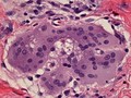
- Digital Images
- Online
Multinucleated giant cell containing an asteroid, microscopy.
William R. Geddie
- Digital Images
- Online
The use of analine dyes in microscopy, Paul Ehrlich

- Digital Images
- Online
Streptococci gordonii biofilm grown on a dental restorative; imaged by scanning electron microscopy.
Gemma Cotton
- Digital Images
- Online
Confocal micrograph of Bacillus subtilis. Bacillus subtilis is a Gram-positive, rod-shaped bacterium, commonly found in soil. Fluorescent proteins (TagRFP-T, sfGFP, TagBFP, mKate2 and mOrange2), time-lapse confocal microscopy and biophysical models are being used to understand the organization of bacterial biofilms.
Fernan Federici & Jim Haseloff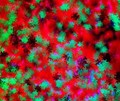
- Digital Images
- Online
Vitamin C (ascorbic acid) crystals imaged by cross polarised light microscopy. Vitamin C is an antioxidant and is important for collagen formation and wound healing. A good source of vitamin C is found in a variety of fruit and vegetables including citrus friuts, brussels sprouts and broccoli. It is a water soluble vitamin that cannot be stored in the body so needs to be ingested regularly. A lack of Vitamin C causes scurvy. 100X image magnification.
Kevin Mackenzie, University of Aberdeen
- Digital Images
- Online
Fibroblast cells with endocytosed particles
Alex Gray
- Digital Images
- Online
Fibroblast cells with endocytosed particles
Alex Gray
- Digital Images
- Online
Fibroblast cells with endocytosed particles
Alex Gray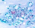
- Digital Images
- Online
Papanicolaou stained smear of a clival chordoma, microscopy. Chordomas are cancers formed of cells which resemble those of the notochord (spine) of a developing foetus. Although they can present anywhere within the spine and skull, the majority grow in the sacral region of the spine, corresponding to the lower back. This image shows a Papanicolaou (Pap) stained smear obtained from a needle biopsy of a chordoma in the clivus, a part of the cranium at the base of the skull.
William R. Geddie