209 results filtered with: Digital Images, Pictures
- Pictures
Microscopy: diagrams illustrating insects and parts of insects. Engraving by A. Bell.
Reference: 46494i- Pictures
Microscopy: diagrams illustrating insects and parts of insects. Engraving by A. Bell.
Reference: 46496i
- Pictures
- Online
Microscopy: diagrams illustrating insects and parts of insects. Engraving by A. Bell.
Reference: 46489i- Pictures
Microscopy: diagrams illustrating insects and parts of insects. Engraving by A. Bell.
Reference: 46498i- Pictures
Microscopy: diagrams illustrating insects and parts of insects. Engraving by A. Bell.
Reference: 46497i- Pictures
Microscopy: diagrams illustrating insects and parts of insects. Engraving by A. Bell.
Reference: 46495i
- Pictures
- Online
Microscopy: diagrams illustrating insects and parts of insects. Engraving by A. Bell.
Reference: 46487i
- Pictures
- Online
Microscopy: diagrams illustrating insects and parts of insects. Engraving by A. Bell.
Reference: 46485i
- Pictures
- Online
Microscopy: diagrams illustrating insects and parts of insects. Engraving by A. Bell.
Reference: 46486i
- Pictures
- Online
Microscopy: diagrams illustrating insects and parts of insects. Engraving by A. Bell.
Reference: 46488i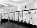
- Digital Images
- Online
The Microscopy and Zoology.

- Digital Images
- Online
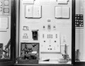
The Microscopy and Zoology.
- Digital Images
- Online
The Microscopy and Zoology.

- Digital Images
- Online
Myxoid liposarcoma, microscopy
William R. Geddie
- Digital Images
- Online
Human kidney cell, Gated-STED microscopy
Alison Dun, ESRIC (Edinburgh Super-Resolution Imaging Consortium)
- Digital Images
- Online
COS-7 cell, confocal and supre resolution microscopy
Daniela Malide, NIH, Bethesda, USA
- Digital Images
- Online
Squamous carcinoma of lacrimal gland, microscopy
William R. Geddie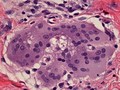
- Digital Images
- Online
Multinucleated giant cell containing an asteroid, microscopy.
William R. Geddie
- Digital Images
- Online
The use of analine dyes in microscopy, Paul Ehrlich

- Digital Images
- Online
Streptococci gordonii biofilm grown on a dental restorative; imaged by scanning electron microscopy.
Gemma Cotton
- Pictures
- Online
Optics: microscopy, including a magnified title page and a specimen holder. Engraving by Barlow.
Reference: 47507i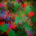
- Digital Images
- Online
Confocal micrograph of Bacillus subtilis. Bacillus subtilis is a Gram-positive, rod-shaped bacterium, commonly found in soil. Fluorescent proteins (TagRFP-T, sfGFP, TagBFP, mKate2 and mOrange2), time-lapse confocal microscopy and biophysical models are being used to understand the organization of bacterial biofilms.
Fernan Federici & Jim Haseloff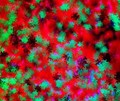