65 results
- Books
Back-bone : photographed from "The Scalpel" / by Edward H. Dixon.
Dixon, Edward H., 1808-1880.Date: [1866]- Books
Clever backbone / John Agard.
Agard, John, 1949-Date: 2009
- Archives and manuscripts
- Online
Diagram referenced as "DNA base sugar phosphate "backbone""
Miss JacksonDate: March 1956Reference: KDBP/1/1/1900Part of: King's College London Department of Biophysics
- Digital Images
- Online
Illustration of the DNA double helix. The sugar-phosphate backbone of the two complementary strands are visible (red and blue).
Susan Lockhart- Books
The backbone of history : health and nutrition in the Western hemisphere / edited by Richard H. Steckel and Jerome C. Rose.
Date: 2002- Books
Animals without backbones : an introduction to the invertebrates / Ralph Buchsbaum.
Buchsbaum, Ralph, 1907-2002.Date: 1951- Books
Animals in Saxon & Scandinavian England : backbones of economy and society / Matilida Holmes.
Holmes, MatildaDate: [2014]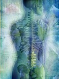
- Digital Images
- Online
Back pain
Bill McConkey
- Pictures
- Online
A skeleton of a child born with two backbones and two heads. Collotype by Römmler & Jonas after a radiograph made for G. Leopold and Th. Leisewitz, 1908.
Leopold, G. (Gerhard), 1846-1911.Date: 1908Reference: 17138i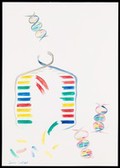
- Digital Images
- Online
Illustration depicting semi-conservative DNA replication. A DNA double helix prior to replication is shown in the top left of the image. The sugar phosphate backbone and nucleotide bases are visible. Complementary base pairing of adenine with thymine (blue with green) and guanine with cytosine (red with yellow) is shown. During replication, a length of the double helix temporarily unwinds and separates into two strands. Free nucleotides bind by complementary base pairing to the recently exposed nucleotides on each strand which act as a template. Two new double helices are formed, each containing one original generation and one new generation strand of DNA. The sequence of base pairs in each double helix is identical to the original.
Susan Lockhart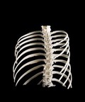
- Digital Images
- Online
Ribcage, Hodgkin lymphoma patient, 3D printed nylon
Dave Farnham
- Digital Images
- Online
Ribcage, Hodgkin lymphoma patient, 3D printed nylon
Dave Farnham
- Digital Images
- Online
Ribcage, Hodgkin lymphoma patient, 3D printed nylon
Dave Farnham
- Digital Images
- Online
Ribcage, Hodgkin lymphoma patient, 3D printed nylon
Dave Farnham
- Digital Images
- Online
Ribcage, Hodgkin lymphoma patient, 3D printed nylon
Dave Farnham
- Digital Images
- Online
Mouse embryo
Macroscopic Solutions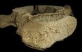
- Digital Images
- Online
Single human T8 vertebra from infant at term, micro-CT
Frank Acquaah
- Digital Images
- Online
Mouse embryo
Macroscopic Solutions
- Digital Images
- Online
Ribcage, Hodgkin lymphoma patient, 3D printed nylon
Dave Farnham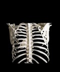
- Digital Images
- Online
Heart in ribcage, Hodgkin lymphoma patient, 3D printed nylon
Dave Farnham
- Digital Images
- Online
Heart in ribcage, Hodgkin lymphoma patient, 3D printed nylon
Dave Farnham
- Digital Images
- Online
Heart in ribcage, Hodgkin lymphoma patient, 3D printed nylon
Dave Farnham
- Digital Images
- Online
Heart in ribcage, Hodgkin lymphoma patient, 3D printed nylon
Dave Farnham
- Digital Images
- Online
Heart in ribcage, Hodgkin lymphoma patient, 3D printed nylon
Dave Farnham
- Digital Images
- Online
Heart in ribcage, Hodgkin lymphoma patient, 3D printed nylon
Dave Farnham