514 results filtered with: Pictures, Digital Images
- Pictures
A Chinese man displaying symptoms of anthrax of the artery of the neck. Coloured line block print by Chiang Yee, after a Chinese artist, 1920/1940?.
Date: [1920/1940?]Reference: 573690i- Pictures
Meningioma in a 78-year old man with a brain tumour and fatal pulmonary artery thrombosis. Watercolour by Barbara E. Nicholson, 1951.
Nicholson, BarbaraDate: 1951Reference: 33998iPart of: Barbara Nicholson medical illustration collection.
- Pictures
- Online
Ambroise Paré using a ligature on an artery of an amputated leg of a soldier, during the Siege of Metz, 1553. Photogravure after T. Chartran, 1889.
Chartran, Théobald, 1849-1907.Date: 1889Reference: 22690i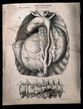
- Pictures
- Online
A diseased artery (aneurism of the aorta); and a diseased section of bone, numbered for key. Lithograph by Batelli after Ottavio Muzzi, c. 1843.
Muzzi, Ottavio.Reference: 34144i- Pictures
Heart section, with ruptured artery causing compression and infiltration in a 71-year old woman with cardiac tamponade. Watercolour by Barbara E. Nicholson, 1947.
Nicholson, BarbaraDate: 1947Reference: 32216iPart of: Barbara Nicholson medical illustration collection.- Pictures
Surgery: surgical instruments, including instruments to compress an artery (tourniquet), compresses and bands. Engraving with etching by A.J. Defehrt after L.-J. Goussier.
Goussier, Louis-Jacques, 1722-1799.Reference: 47635i- Pictures
Fatal obstructive jaundice in a female patient with atresia of bile ducts: greatly hypertrophied hepatic artery, microscopic view. Watercolour by Barbara E. Nicholson, 1956.
Nicholson, BarbaraDate: 1956Reference: 35641iPart of: Barbara Nicholson medical illustration collection.
- Pictures
- Online
Internal organs and arteries of the horse: five figures showing dissections of the heart, lungs, thoracic viscera, pulmonary artery and veins. Engraving by T. Cowan after B. Herring, ca. 1860.
Herring, Benjamin, 1830-1871.Date: [1860?]Reference: 570542i- Pictures
Three sections of an aneurysm, bulging at the aortic arch, in the aorta and branching artery in an 69-year old man. Watercolour by Barbara E. Nicholson, 1947.
Nicholson, BarbaraDate: 1947Reference: 32105iPart of: Barbara Nicholson medical illustration collection.
- Pictures
Blood circulation in the respiratory system, showing the pulmonary artery and vein. Ink and white paint on paper, with acetate overlay, by D. Vaihinger, 1975.
Vaihinger, Dagmar.Date: 1975Reference: 569717i
- Pictures
Ambroise Paré, on the battlefield using a ligature for the artery of an amputated leg of a soldier. Wood engraving by Charles Maurand after E. Morin.
Morin, Edmond, 1824-1882.Date: 1800-1899Reference: 22688i- Pictures
Heart with obstructed descending branch of coronary artery, seen split into five sections in a 52-year old woman with coronary thrombosis and infarct. Watercolour by Barbara E. Nicholson, 1948.
Nicholson, BarbaraDate: 1948Reference: 32675iPart of: Barbara Nicholson medical illustration collection.- Pictures
Cardiac infarction and thrombosis in a 52-year old man with hemiplegia: heart sections and inset detail of clot in descending left coronary artery. Watercolour by Barbara E. Nicholson, 1953.
Nicholson, BarbaraDate: 1953Reference: 35015iPart of: Barbara Nicholson medical illustration collection.- Pictures
Pulmonary thrombosis in a male patient with fatal pneumonia and cyanosis: detail of right pulmonary artery in section, completely occluded by clot, which has also spread to left arteries. Watercolour by Barbara E. Nicholson, 1954.
Nicholson, BarbaraDate: 1954Reference: 35123iPart of: Barbara Nicholson medical illustration collection.
- Pictures
- Online
Underside of a brain, shown beside a dissection of the abdominal aorta and left iliac artery, both showing symptoms of atherosclerosis and aneurysm. Chromolithograph by W. Gummelt, ca. 1897.
Gummelt, W.Date: [1897?]Reference: 577230i- Pictures
A ruptured aneurysm from the carotid artery, lying against the pituitary gland in a 70-year old man with terminal haemorrhaging following rupture. Watercolour by Barbara E. Nicholson, 1947.
Nicholson, BarbaraDate: 1947Reference: 32106iPart of: Barbara Nicholson medical illustration collection.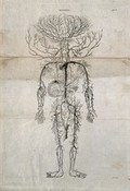
- Pictures
- Online
Human arterial system. Engraving by Campbell, after an engraving by M. Vandergucht after W. Cowper, for Drake, 1707.
Cowper, William, 1666-1709.Reference: 37384i- Pictures
Large left hydronephrosis with multiple opaque renal calculi in a 47-year old man with urinary infection: detail showing aneurysm occluding mesenteric artery supply. Watercolour by Barbara E. Nicholson, 1950.
Nicholson, BarbaraDate: 1950Reference: 33854iPart of: Barbara Nicholson medical illustration collection.- Pictures
Section of heart, with detail, showing distortion due to large clot in right pulmonary artery, with interventrical defects in a 43-year old woman with pleurisy and congestive heart failure. Watercolour by Barbara E. Nicholson, 1948.
Nicholson, BarbaraDate: 1948Reference: 32478iPart of: Barbara Nicholson medical illustration collection.- Pictures
Posterior view of thrombus in the left pulmonary artery and distorted lung with inter-ventrical defects in a 43-year old woman with pleurisy and congestive heart failure. Watercolour by Barbara E. Nicholson, 1948.
Nicholson, BarbaraDate: 1948Reference: 32481iPart of: Barbara Nicholson medical illustration collection.- Pictures
Section of main mesenteric artery, with structural decomposition and invasion of cavity, showing thrombus attached to the fold of a growth, in a male patient with fatal thrombosis and inflammation of the arteries. Watercolour by Barbara E. Nicholson, 1947.
Nicholson, BarbaraDate: 1947Reference: 32176iPart of: Barbara Nicholson medical illustration collection.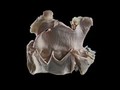
- Digital Images
- Online
Aortic valvular endocarditis, horse. This equine (horse) aorta has been dissected at the level of the valve, separating the artery from the left ventricle (bottom chamber). Nodules are present which have formed as a consequence of endocarditis.
Michael Frank, Royal Veterinary College- Pictures
Dissecting aneurysm of aorta in a male patient: section showing (a) aortic valve and extensively diseased descending aorta cloaked in large blood clot, with (b) inset of independant thrombus in carotid artery. Watercolour by Barbara E. Nicholson, 1958.
Nicholson, BarbaraDate: 1958Reference: 36031iPart of: Barbara Nicholson medical illustration collection.- Pictures
Anterior and posterior walls of consuming gastric ulcer in stomach when opened, turned back and drawn in section, in a 55-year old man with fatal haemorrhage (due to artery eroded by ulcer). Watercolour and black ink diagram by Barbara E. Nicholson, 1948.
Nicholson, BarbaraDate: 1948Reference: 32676iPart of: Barbara Nicholson medical illustration collection.- Pictures
Congestive heart failure in a 70-year old man with digitalis, dyspnoea and chronic abdominal distension: surgical specimens of (a) embolism in superior mesenteric artery, (b) intra-mural thrombus in left ventricle and (c) diagram of gangrenous small intestine. Watercolour by Barbara E. Nicholson, 1951.
Nicholson, BarbaraDate: 1951Reference: 34211iPart of: Barbara Nicholson medical illustration collection.