1,787 results

- Digital Images
- Online
Cell walls in a Quercus (oak) stem, LM
Fernán Federici- Books
p16INK4A as a marker for cervical dyskaryosis / Niamh Murphy, Cynthia Heffron, Martina Ring, Orla Sheils, John J. O'Leary, The Coombe Women's Hospital, St. James's Hospital and Trinity College Dublin.
Date: 2004
- Digital Images
- Online
Drosophila ovaries stained for actin/purple and DNA/orange.
Teresa Niccoli & Daniel St Johnston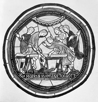
- Archives and manuscripts
- Online
M0003548: Stained glass window at Canterbury
Date: 5 July 1933Reference: WT/D/1/20/1/29/28Part of: Wellcome Trust Corporate Archive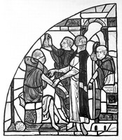
- Archives and manuscripts
- Online
M0003547: Stained glass window at Canterbury
Date: 5 July 1933Reference: WT/D/1/20/1/29/27Part of: Wellcome Trust Corporate Archive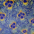
- Digital Images
- Online
Cross-section through a bamboo stem, LM
Fernán Federici
- Digital Images
- Online
Fibroblasts stained to show viability
Biosciences Imaging Gp, Soton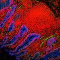
- Digital Images
- Online
Human small intestine showing villi and glands. The cytokeratinin the cells is stained blue, the cell nuclei are stained red and the endothelial cells lining the blood vessels are stained green.
S. Schuller
- Digital Images
- Online
Human small intestine showing villi and glands. The cytokeratinin the cells is stained blue, the cell nuclei are stained red and the endothelial cells lining the blood vessels are stained green.
S. Schuller
- Digital Images
- Online
Human small intestine showing villi and glands. The cytokeratinin the cells is stained blue, the cell nuclei are stained red and the endothelial cells lining the blood vessels are stained green.
S. Schuller
- Digital Images
- Online
Human small intestine showing villi. The cytokeratinin the cells is stained blue, the cell nuclei are stained red and the endothelial cells lining the blood vessels are stained green.
S. Schuller
- Digital Images
- Online
Xenopus cancer kidney cells, prometaphase
Paul Andrews/Univ. Dundee
- Digital Images
- Online
Xenopus cancer kidney cells,telophase
Paul Andrews/Univ. Dundee
- Digital Images
- Online
Human small intestine showing villi and glands. The cytokeratin in the cells is stained blue, the cell nuclei are stained red and the endothelial cells lining the blood vessels are stained green.
S. Schuller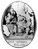
- Archives and manuscripts
- Online
M0004441: Amputation of a leg, stained glass fragment
Date: 13 November 1935Reference: WT/D/1/20/1/35/64Part of: Wellcome Trust Corporate Archive- Books
Khusra : stains & stencils / Qasim Riza Shaheen.
Shaheen, Qasim RizaDate: 2007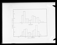
- Archives and manuscripts
- Online
"Histogram of Feulgen stain. Linear and log scales."
Walker, Peter M. B.Date: November 1952Reference: KDBP/1/1/0729Part of: King's College London Department of Biophysics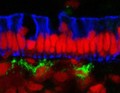
- Digital Images
- Online
Human small intestine showing the columnar epithelium. The cytokeratinin the cells is stained blue, the cell nuclei are stained red and the endothelial cells lining the blood vessels are stained green.
S. Schuller- Archives and manuscripts
Examples only, stained sections and x-rays
Date: c.1920s-1940sReference: PP/MEL/C/71Part of: Mellanby, Sir Edward- Archives and manuscripts
- Online
TEM Prints on Phage - Negative Staining
Date: 1956Reference: SB/6/2/4Part of: Sydney Brenner Collection
- Digital Images
- Online
Multi-sized beads (constellation stained), fluorescence
Kevin Mackenzie, University of Aberdeen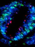
- Digital Images
- Online
Section through neural tube of chick embryo, confocal
Arwen Wilcock/Univ. Dundee
- Digital Images
- Online
Cytokinesis in neural tube cells of chick embryo
Arwen Wilcock/Univ. of Dundee
- Digital Images
- Online
Cytokinesis in neural tube cells of chick embryo
Arwen Wilcock / Univ. of Dundee
- Digital Images
- Online
Outer hair cells stained for actin
Dr David Furness