722 results

- Digital Images
- Online
Balanced reciprocal translocation 46,XY,t(2;5). This male has a chromosomal disorder. A chromosome 2 and a chromosome 5 have exchanged segments. The cell still contains a complete complement of
Wessex Reg. Genetics Centre
- Digital Images
- Online
Human T cells showing nuclei
A. Walker, L. Sharp & J. Pryde- Books
Cost-project 83-84, production and feeding of single cell protein : first annual report 1980-81 / Commission of the European Communities.
Date: 1981
- Digital Images
- Online
Cellular architecture of normal human skin imaged by whole mount tissue microscopy. Human skin has a rich network of white blood cells (specifically dendritic cells, T cells and macrophages) which form sheaths around blood vessels. In this image, T cells (stained for CD3; red) dendritic cells (stained for MHC class II; green) and macrophages (stained for LYVE-1; blue with some cells showing a tinge of green) can be seen. Cell nuclei have been stained with DAPI (grey). This normal cellular architecture is grossly disrupted in diseased skin (see related images). X10 magnification. Scale bar (white) represents 200 micrometres.
Dr. Xiao-nong Wang, Human Dendritic Cell Laboratory, Newcastle University
- Digital Images
- Online
Cellular architecture of normal human skin imaged by whole mount tissue microscopy. Human skin has a rich network of white blood cells (specifically dendritic cells, T cells and macrophages) which form sheaths around blood vessels. In this image, T cells (stained for CD3; red) dendritic cells (stained for MHC class II; green) and macrophages (stained for LYVE-1; blue with some cells showing a tinge of green) can be seen. Cell nuclei have been stained with DAPI (grey). This normal cellular architecture is grossly disrupted in diseased skin (see related images). X20 magnification. Scale bar (white) represents 100 micrometres.
Dr. Xiao-nong Wang, Human Dendritic Cell Laboratory, Newcastle University
- Digital Images
- Online
Human embryonic stem cells
Annie Cavanagh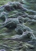
- Digital Images
- Online
Human embryonic stem cells
Annie Cavanagh
- Digital Images
- Online
Human embryonic stem cells
Annie Cavanagh
- Digital Images
- Online
Cellular architecture of normal human skin imaged by whole mount tissue microscopy. Human skin has a rich network of white blood cells (specifically dendritic cells, T cells and macrophages) which form sheaths around blood vessels. In this image, blood vessels (string-like structures stained for CD31; green), lymphatic vessels (ribbon-like structures stained for LYVE-1; blue) and T cells (stained for CD3; red) can be seen. T cells are only found around dermal blood vessels. Macrophages (stained for LYVE-1; blue) are also present. This normal cellular architecture is grossly disrupted in diseased skin (see related images). X10 magnification. Scale bar (white) represents 200 micrometres.
Dr. Xiao-nong Wang, Human Dendritic Cell Laboratory, Newcastle University- Videos
Interference microscopy of normal and cancer cells in tissue culture.
Date: 2015- Books
Cell death / edited by Lawrence M. Schwartz and Barbara A. Osborne.
Date: [1995], ©1995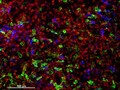
- Digital Images
- Online
Cellular architecture of human skin lymphoma imaged by whole mount tissue microscopy. Normal human skin has a rich network of white blood cells (specifically dendritic cells, T cells and macrophages) which form sheaths around blood vessels. In diseased skin, such as in skin lymphoma as seen here, this normal architecture becomes distorted. In this image, lots of T cells (stained for CD3; red), dendritic cells (stained for CD11c; green) and macrophages (stained for LYVE-1; blue) have infiltrated the skin. X20 magnification. Scale bar (white) represents 100 micrometres.
Dr. Xiao-nong Wang, Human Dendritic Cell Laboratory, Newcastle University- Film
Interference microscopy of normal and cancer cells in tissue culture.
Date: 1958- Videos
- Online
Interference microscopy of normal and cancer cells in tissue culture.
- Books
Monoclonal T cells and their products / edited by Marc Feldmann, Max H. Schreier.
Date: 1982
- Digital Images
- Online
Cellular architecture of normal human skin imaged by whole mount tissue microscopy. Human skin has a rich network of white blood cells (specifically dendritic cells, T cells and macrophages) which form sheaths around blood vessels. In this image, blood vessels (string-like structures stained for CD31; red), lymphatic vessels (ribbon-like structures stained for LYVE-1; blue) and dendritic cells (stained for CD11c; green) can be seen. Macrophages (stained for LYVE-1; blue) are also present. This normal cellular architecture is grossly disrupted in diseased skin (see related images). X10 magnification. Scale bar (white) represents 200 micrometres.
Dr. Xiao-nong Wang, Human Dendritic Cell Laboratory, Newcastle University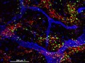
- Digital Images
- Online
Cellular architecture of normal human skin imaged by whole mount tissue microscopy. Human skin has a rich network of white blood cells (specifically dendritic cells, T cells and macrophages) which form sheaths around blood vessels (string-like structures). A network of lymphatic vessels (ribbon-like structures) is also present. In this image, human skin lymphatic vessels (stained for LYVE-1; blue) and white blood cells comprised of dendritic cells (stained for CD11c; green) and T cells (stained for CD3; red) can be seen. Some macrophages also express the protein LYVE-1 similar to lymphatic vessel cells which can be appreciated as blue cells within and in between the sheaths of white blood cells. This normal cellular architecture is grossly disrupted in diseased skin (see related images). X10 magnification. Scale bar (white) represents 200 micrometres.
Dr. Xiao-nong Wang, Human Dendritic Cell Laboratory, Newcastle University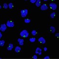
- Digital Images
- Online
Human T cells showing nuclei & Golgi bodies
A. Walker, L. Sharp & J. Pryde
- Books
- Online
Origin : Cancer cell of sarcoma one; identical in Cytology. Finding the pure cancer Cytology, only, in a reported case of undifferentiated round cell sarcoma. Revision of many cases of sarcoma yield pure carcinoma on restudy. Does 'cure of cancer' 10-18 years, post operation with no recurrences, then fatal recurrences, suggest a dormant abeyant chronic form of cancer- A benign form of Cancer? Various (30) cases in point of discussion. The causal diminutive cancer cellulettes, dormant or active, must be neutralized or destroyed for ultimate cure. A further amplification of the specific cancer cellule and its conduct. Transmutation. Cell division. / by Frank A. Stahl, M.D.
Stahl, Frank August, 1862-Date: 1887- Books
Health economics, management & policy / edited by John N. Yfantopoulos & Gregory T. Papanikos.
Date: 2005
- Digital Images
- Online
Cellular architecture of normal human skin imaged by whole mount tissue microscopy. Human skin has a rich network of white blood cells (specifically dendritic cells, T cells and macrophages) which form sheaths around blood vessels. This image was taken greater than 150 micrometres beneath the junction that joins the dermal and epidermal layers of the skin (dermo-epidermal junction). At this level, dendritic cells (stained for CD11c; green) and macrophages (stained for LYVE-1; blue) form clusters around blood vessels (stained for CD31; red). This normal cellular architecture is grossly disrupted in diseased skin (see related images). Scale bar (white) represents 100 micrometres.
Dr. Xiao-nong Wang, Human Dendritic Cell Laboratory, Newcastle University
- Digital Images
- Online
Cellular architecture of normal human skin imaged by whole mount tissue microscopy. Human skin has a rich network of white blood cells (specifically dendritic cells, T cells and macrophages) which form sheaths around blood vessels. This image was taken less than 20 micrometres beneath the junction that joins the dermal and epidermal layers of the skin (dermo-epidermal junction). At this level, dendritic cells (stained for CD11c; green) form clusters around and between blood capillary loops (stained for CD31; red). The blind-ended tips of initial lymphatic vessels are just visible (stained for LYVE-1; blue) at this level. This normal cellular architecture is grossly disrupted in diseased skin (see related images). Scale bar (white) represents 200 micrometres.
Dr. Xiao-nong Wang, Human Dendritic Cell Laboratory, Newcastle University- Videos
Don't die young. Series 1, part 5, Skin.
Date: 2007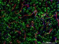
- Digital Images
- Online
Cellular architecture of normal human skin imaged by whole mount tissue microscopy. Human skin has a rich network of white blood cells (specifically dendritic cells, T cells and macrophages) which form sheaths around blood vessels. This image was taken directly beneath the junction that joins the dermal and epidermal layers of the skin (dermo-epidermal junction). At this level, the capillary network (stained for CD31; red) is visualised against a lawn of autofluorescent dermal papillae (finger-like projections of the dermis; green) scattered with dendritic cells (stained for CD11c; green) and macrophages (stained for LYVE-1; blue). This normal cellular architecture is grossly disrupted in diseased skin (see related images). Scale bar (white) represents 200 micrometres.
Dr. Xiao-nong Wang, Human Dendritic Cell Laboratory, Newcastle University- Books
Apoptosis / edited by Lawrence M. Schwartz, Jonathan D. Ashwell.
Date: [2001], ©2001