Wellcome uses cookies.
Read our policy
Close cookie notification
Skip to main content
Wellcome Collection homepage
Visit us
What’s on
Stories
Collections
Get involved
About us
Sign in to your library account
Search our stories, images, catalogue and events
Library account
Search our stories, images, catalogue and events
Search
Images search
Search for images
Search
All
Stories
Images
Catalogue
Events
Colours
Licences
Creative Commons CC-BY (70)
Creative Commons CC-BY-NC (17)
Creative Commons CC0 (13)
In copyright (12)
Public Domain Mark (11)
Dates
From
to
Types/Techniques
Leaflets (10)
Ephemera (9)
Engravings (6)
Price lists (6)
Lithographs (4)
Portrait prints (4)
Intaglio prints (3)
Posters (3)
Etchings (2)
Broadsides (1)
Caricatures (1)
Landscape prints (1)
Photomechanical prints (1)
Prints (1)
Subjects
T cell (37)
Blue (26)
Green (24)
Red (23)
Immune response (16)
AIDS (14)
Pattern (14)
Purple (14)
Yellow (14)
Cancer (12)
Shape (12)
WHITE BLOOD CELLS (12)
AFFINITY MATURATION (11)
Immunology (11)
IMMUNOLOGY (11)
Lymphocyte (11)
Retrovirus (11)
Virus (11)
ANTIGEN RECEPTOR (10)
B cell (10)
Contributors
Peter Lane and Fiona McConnell (12)
Dr. Xiao-nong Wang, Human Dendritic Cell Laboratory, Newcastle University (9)
David S. Goodsell, The Scripps Research Institute (8)
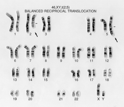
Wessex Reg. Genetics Centre (8)
Odra Noel (7)
AGMED Inc. (Bedford, MA.) (6)
Kate Whitley (5)
T.Blundell & N Campillo (5)
Fernán Federici (4)
K R Acharya (4)
Annie Cavanagh (3)
Emei Ma, P/C Guy McLoughlin (3)
Steve Winder (3)
WAK-Chemie Medical (Firm) (3)
A. Walker, L. Sharp & J. Pryde (2)
Amsler, Samuel, 1791-1849 (2)
Dr Jeremy Skepper (2)
Kaulbach, Wilhelm von, 1804-1874 (2)
Langlois, François, 1588-1647 (2)
Le Bas, Jacques-Philippe, 1707-1783 (2)
Submit
125 results
Search result sorting
Sort by:
Relevance
Production dates
Sort order:
Ascending
Descending
Submit
Page
1
of 5
Next (page 2)
Close modal window
Page
1
of 5
Next (page 2)


![Turn this column upside down : Uni-Sorb ready-to-use nylon wool columns for the preparation fo [sic] enriched T and B cell fractions / WAK-Chemie Medical GmbH.](https://iiif.wellcomecollection.org/image/b15647468_EPH_28_8_0001.jp2/full/282%2C/0/default.jpg)