74 results filtered with: Pictures
- Pictures
Microscopy: parts of a louse. Engraving [after R. Hooke?].
Reference: 46518i- Pictures
Microscopy: fresh water melicerta. Coloured lithograph by L. Aldous.
Reference: 46477i- Pictures
Microscopy: fresh water rotifers. Coloured lithograph by L. Aldous.
Reference: 46484i- Pictures
Microscopy: parts of a ladybird. Engraving [after R. Hooke?].
Hooke, Robert, 1635-1703.Reference: 46523i
- Pictures
- Online
Microscopy: parts of a louse [?]. Engraving [after R. Hooke?].
Hooke, Robert, 1635-1703.Reference: 46519i- Pictures
Microscopy: parts of a plant. Engraving [after A. van Leeuwenhoek?].
Reference: 46511i- Pictures
Microscopy: parts of a plant [?]. Engraving [after R. Hooke?].
Hooke, Robert, 1635-1703.Reference: 46524i- Pictures
Microscopy: fresh water monads and stentors. Coloured lithograph by L. Aldous.
Reference: 46474i- Pictures
Microscopy: parts of a seed-pod [?]. Engraving [after A. van Leeuwenhoek?].
Reference: 46509i- Pictures
Microscopy: parts of a louse [or flea?]. Engraving [after A. van Leeuwenhoek?].
Reference: 46512i
- Pictures
- Online
Optics: microscopy, including a magnified title page and a specimen holder. Engraving by Barlow.
Reference: 47507i
- Pictures
- Online
The liver and the kidneys. Engraving, 1686, the third, fourth and fifth figures after G. de Lairesse, 1685.
Lairesse, Gérard de, 1640-1711.Date: [1686]Reference: 31500i
- Pictures
- Online
Left, a woman personifying science instructing her children in how to use a microscope; a woman personifying truth reveals the scene to Father Time. Mezzotint after T.S. Duché, 1787.
Duché, Thomas Spence, 1763-1790.Date: July 1st 1787Reference: 577912i
- Pictures
- Online
The skin, in microscopic view (figures 1-2), and muscles (figures 3-6). Engraving, 1686.
Date: [1686]Reference: 31568i
- Pictures
- Online
A human skeleton, seen from the front and leaning on a spade, surrounded by illustrations of individual bones. Engraving, 1686.
Date: [1686]Reference: 32187i
- Pictures
- Online
The breast dissected and the nipple and areola and a hair as viewed under a microscope. Engraving, 1686.
Date: [1686]Reference: 31565i
- Pictures
- Online
The stomach, peritoneum and oesophagus. Engraving, 1686, after Gérard de Lairesse, 1685.
Lairesse, Gérard de, 1640-1711.Date: [1686]Reference: 30014i
- Pictures
- Online
A pink microscope reflecting a magnified view of the HIV virus against a black and red background with Maldivian (Divehi) lettering; an advertisement for safe sex to prevent AIDS. Colour lithograph, ca. 1996.
Date: [1996?]Reference: 677662i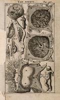
- Pictures
- Online
A foetus and placenta with umbilical cord. Engraving, 1686, the second, third, fifth and sixth figures after G. de Lairesse, 1685.
Lairesse, Gérard de, 1640-1711.Date: [1686]Reference: 31559i
- Pictures
A physician telling a patient that he is going to die: the patient stares out at the viewer. Colour photogravure after the Hon. J. Collier, 1908.
Collier, John, 1850-1934.Date: [1908]Reference: 21878i
- Pictures
Albert Nachet, microscopist. Oil painting by Harry Herman Salomon after a photograph.
Salomon, Harry Herman, 1860-1936.Reference: 45760i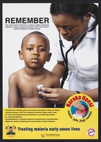
- Pictures
- Online
A nurse uses a stethoscope on a child with a thermometer in his mouth: preventing malaria in Kenya. Colour lithograph by Ministry of Public Health and Sanitation, ca. 2008.
Date: [2008?]Reference: 755556i
- Pictures
- Online
Two enlarged images of T-cells one infected with HIV by the Frederick Cancer Research and Development Center. Colour lithograph by Nancy Burson and Kunio Nagashima, 1991.
Date: 1991Reference: 667046i- Pictures
Chemo Day Drawings
Smith, ClareDate: 2019-