1,019 results filtered with: Digital Images

- Digital Images
- Online
Sri Lankan mask with blue coloured face and tall red hat.

- Digital Images
- Online
Macrophage showing the nucleus in blue and the microtubules in green.
NIMR, Francis Crick Institute
- Digital Images
- Online
A blue horse taken from a Persian manuscript on the natural sciences in Nasta'liq script
- Digital Images
- Online
Normal mouse colon showing nuclei in red and the actin in the muscle layer in blue.
S. Schuller
- Digital Images
- Online
An X-ray valve tube, common in blue glass. Inscribed 'Made in England by Cuthbert Andrews'
Cuthbert Andrews
- Digital Images
- Online
Mexican funerary urn of pottery with polychrome decoration. Standing figure of a god with Ocelot headress wearing blue jewel on breast.

- Digital Images
- Online
Human cell in interphase showing the tubulin component of the cytoskeleton in green, the DNA in blue and the kinetochores in pink.
Matthew Daniels
- Digital Images
- Online
Blood vessels in the retina showing the endothelial cells in red and the vascular contents in green. Surrounding cell nuclei are stained blue.
Freya Mowat
- Digital Images
- Online
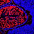
Human small intestine showing villi. The cytokeratinin the cells is stained blue, the cell nuclei are stained red and the endothelial cells lining the blood vessels are stained green.
S. Schuller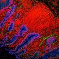
- Digital Images
- Online
Human small intestine showing villi and glands. The cytokeratinin the cells is stained blue, the cell nuclei are stained red and the endothelial cells lining the blood vessels are stained green.
S. Schuller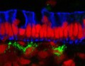
- Digital Images
- Online
Human small intestine showing the columnar epithelium. The cytokeratinin the cells is stained blue, the cell nuclei are stained red and the endothelial cells lining the blood vessels are stained green.
S. Schuller
- Digital Images
- Online
Human small intestine showing villi and glands. The cytokeratinin the cells is stained blue, the cell nuclei are stained red and the endothelial cells lining the blood vessels are stained green.
S. Schuller
- Digital Images
- Online
Human small intestine showing villi and glands. The cytokeratinin the cells is stained blue, the cell nuclei are stained red and the endothelial cells lining the blood vessels are stained green.
S. Schuller
- Digital Images
- Online
Human small intestine showing villi and glands. The cytokeratin in the cells is stained blue, the cell nuclei are stained red and the endothelial cells lining the blood vessels are stained green.
S. Schuller
- Digital Images
- Online
Pilgrim's token, a blue bottle showing a little chapel. Formerly contained St. Wolfgang's water. The bottle was covered with water-paint in order to show the design, when the lantern slide was made.
- Digital Images
- Online
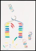
Illustration depicting semi-conservative DNA replication. A DNA double helix prior to replication is shown in the top left of the image. The sugar phosphate backbone and nucleotide bases are visible. Complementary base pairing of adenine with thymine (blue with green) and guanine with cytosine (red with yellow) is shown. During replication, a length of the double helix temporarily unwinds and separates into two strands. Free nucleotides bind by complementary base pairing to the recently exposed nucleotides on each strand which act as a template. Two new double helices are formed, each containing one original generation and one new generation strand of DNA. The sequence of base pairs in each double helix is identical to the original.
Susan Lockhart
- Digital Images
- Online
Cellular architecture of normal human skin imaged by whole mount tissue microscopy. Human skin has a rich network of white blood cells (specifically dendritic cells, T cells and macrophages) which form sheaths around blood vessels. In this image, blood vessels (string-like structures stained for CD31; red), lymphatic vessels (ribbon-like structures stained for LYVE-1; blue) and dendritic cells (stained for CD11c; green) can be seen. Macrophages (stained for LYVE-1; blue) are also present. This normal cellular architecture is grossly disrupted in diseased skin (see related images). X10 magnification. Scale bar (white) represents 200 micrometres.
Dr. Xiao-nong Wang, Human Dendritic Cell Laboratory, Newcastle University
- Digital Images
- Online
Cellular architecture of normal human skin imaged by whole mount tissue microscopy. Human skin has a rich network of white blood cells (specifically dendritic cells, T cells and macrophages) which form sheaths around blood vessels. In this image, blood vessels (string-like structures stained for CD31; green), lymphatic vessels (ribbon-like structures stained for LYVE-1; blue) and T cells (stained for CD3; red) can be seen. T cells are only found around dermal blood vessels. Macrophages (stained for LYVE-1; blue) are also present. This normal cellular architecture is grossly disrupted in diseased skin (see related images). X10 magnification. Scale bar (white) represents 200 micrometres.
Dr. Xiao-nong Wang, Human Dendritic Cell Laboratory, Newcastle University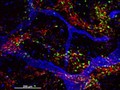
- Digital Images
- Online
Cellular architecture of normal human skin imaged by whole mount tissue microscopy. Human skin has a rich network of white blood cells (specifically dendritic cells, T cells and macrophages) which form sheaths around blood vessels (string-like structures). A network of lymphatic vessels (ribbon-like structures) is also present. In this image, human skin lymphatic vessels (stained for LYVE-1; blue) and white blood cells comprised of dendritic cells (stained for CD11c; green) and T cells (stained for CD3; red) can be seen. Some macrophages also express the protein LYVE-1 similar to lymphatic vessel cells which can be appreciated as blue cells within and in between the sheaths of white blood cells. This normal cellular architecture is grossly disrupted in diseased skin (see related images). X10 magnification. Scale bar (white) represents 200 micrometres.
Dr. Xiao-nong Wang, Human Dendritic Cell Laboratory, Newcastle University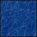
- Digital Images
- Online
Amyloid fibrils, AFM
Dr. Wei-Feng Xue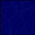
- Digital Images
- Online
Amyloid fibrils, AFM
Dr. Wei-Feng Xue
- Digital Images
- Online
Cellular architecture of normal human skin imaged by whole mount tissue microscopy. Human skin has a rich network of white blood cells (specifically dendritic cells, T cells and macrophages) which form sheaths around blood vessels. In this image, T cells (stained for CD3; red) dendritic cells (stained for MHC class II; green) and macrophages (stained for LYVE-1; blue with some cells showing a tinge of green) can be seen. Cell nuclei have been stained with DAPI (grey). This normal cellular architecture is grossly disrupted in diseased skin (see related images). X10 magnification. Scale bar (white) represents 200 micrometres.
Dr. Xiao-nong Wang, Human Dendritic Cell Laboratory, Newcastle University
- Digital Images
- Online
Cellular architecture of normal human skin imaged by whole mount tissue microscopy. Human skin has a rich network of white blood cells (specifically dendritic cells, T cells and macrophages) which form sheaths around blood vessels. In this image, T cells (stained for CD3; red) dendritic cells (stained for MHC class II; green) and macrophages (stained for LYVE-1; blue with some cells showing a tinge of green) can be seen. Cell nuclei have been stained with DAPI (grey). This normal cellular architecture is grossly disrupted in diseased skin (see related images). X20 magnification. Scale bar (white) represents 100 micrometres.
Dr. Xiao-nong Wang, Human Dendritic Cell Laboratory, Newcastle University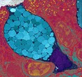
- Digital Images
- Online
Goblet cells are packed full of mucous globules (blue), which they release to provide lubrication and protection to the inner surfaces of the intestine and the respiratory system among others. The mucous globules are condensed inside the goblet cell but expand hugely once they are released, absorbing water within 20 milliseconds. This rapid release occurs in response to lots of different stimuli and allows the mucous to get to work instantly.
University of Edinburgh
- Digital Images
- Online
Cell flower formed from a 3T3 fibroblast cell. An unusually shaped cell found growing under normal conditions. The cell nucleus, containing the DNA, is stained in blue with DAPI. The cell body is stained for F-actin in red to reveal the flowere like shape.
Steve Winder