117 results

- Pictures
- Online
Three diagrams of the organisation of the lobes of the brain for a phrenological textbook. Pen drawing, c. 1902.
Date: 1902Reference: 27967i- Pictures
A brain seen from the underside; with attention to the area associated by Gall with verbal memory. Process print, 1901, after etching, 1809.
Date: 1901Reference: 28011i- Pictures
The brain: side view illustrating the distance of the occipital bone from the phrenological 'organ of philoprogenitiveness'. Process print, 1901, after etching, 1809.
Date: 1901Reference: 28019i
- Pictures
- Online
A male brain, sectioned vertically. Process print, 1901, after etching, 1809.
Date: 1901Reference: 28021i
- Pictures
- Online
A female brain, sectioned vertically: side view. Process print, 1901, after etching, 1809.
Date: 1901Reference: 28022i
- Digital Images
- Online
Human brain activation due to voluntary action
Parashkev Nachev
- Pictures
- Online
The human brain, divided according to Bernard Hollander's system of phrenology. Process print with pen and ink, c. 1902.
Date: [approximately 1902]Reference: 27959i
- Books
- Online
The localisation of cerebral disease : being the Gulstonian lectures of the Royal College of Physicians for 1878 / by David Ferrier.
Date: 1878- Pictures
Two skulls with temporal regions of different size and shape. Photomechanical reproduction with painting, c. 1902.
Date: 1902Reference: 28067i- Pictures
The brain, sectioned vertically; showing the sites of some phrenological faculties. Process print, 1901, after etching, 1809.
Date: 1901Reference: 28020i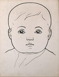
- Pictures
- Online
Head of a child with large cheeks. Drawing, c. 1900.
Date: c. 1900Reference: 28390i- Student dissertations
Henry Charlton Bastian and the cerebral localisation debate / William Brook.
Brook, William.Date: 1989
- Pictures
- Online
Head of woman showing musical ability, according to phrenological classification. Drawing, c. 1900.
Date: c. 1900Reference: 28389i
- Pictures
- Online
Child's head with large temporal lobes and depressed frontal lobe. Drawing, c. 1900.
Date: c. 1900Reference: 28388i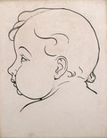
- Pictures
- Online
Child's head, with fat cheeks: profile. Drawing, c. 1900.
Date: c. 1900Reference: 28391i- Pictures
The brain seen from the underside, sectioned horizontally; with attention to the parts associated by Hollander's system of phrenology with the faculties of external perception and its memory. Process print, 1901, after etching, 1809.
Date: 1901Reference: 28018i
- Pictures
- Online
Right profile of head with depressed frontal lobes, divided up to show the location of all the lobes. Drawing, c. 1900.
Date: c. 1900Reference: 28386i
- Pictures
- Online
Left profile of a head showing depressed frontal lobes. Drawing, c. 1900.
Date: c. 1900Reference: 28387i- Film
The mid-brain infant.
Date: 1925 / 1960- Pictures
A skull with a high parietal bone; another indicating diminished frontal and enlarged occipital lobes. 2 photomechanical reproductions, c. 1902.
Date: 1902Reference: 28072i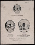
- Pictures
- Online
The bases of three skulls: a new born infant's, a misogynist's, and a man suffering from satyriasis. Process print, 1901, after etching, 1809.
Date: 1901Reference: 28023i- Books
Aux origines du cerveau : localisations, langage et mémoire dans l'oeuvre de Charcot / Jacques Gasser.
Gasser, Jacques.Date: 1995- Pictures
A head divided in two, the left half showing activities numbered 1 to 42. Lithograph by Frank Ellis, 1901.
Ellis, Frank.Date: 1901Reference: 583321i- Videos
Nerves.
Date: 1996
- Books
- Online
The localising value of optic neuritis in intracranial tumour / by J.M. Martin.
Martin, J.M.Date: [1897]