135 results filtered with: Digital Images
- Digital Images
- Online
The Microscopy and Zoology.

- Digital Images
- Online
The Microscopy and Zoology.
- Digital Images
- Online
The Microscopy and Zoology.

- Digital Images
- Online
Myxoid liposarcoma, microscopy
William R. Geddie
- Digital Images
- Online
COS-7 cell, confocal and supre resolution microscopy
Daniela Malide, NIH, Bethesda, USA
- Digital Images
- Online
Human kidney cell, Gated-STED microscopy
Alison Dun, ESRIC (Edinburgh Super-Resolution Imaging Consortium)
- Digital Images
- Online
Squamous carcinoma of lacrimal gland, microscopy
William R. Geddie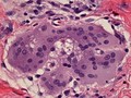
- Digital Images
- Online
Multinucleated giant cell containing an asteroid, microscopy.
William R. Geddie
- Digital Images
- Online
The use of analine dyes in microscopy, Paul Ehrlich

- Digital Images
- Online
Streptococci gordonii biofilm grown on a dental restorative; imaged by scanning electron microscopy.
Gemma Cotton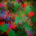
- Digital Images
- Online
Confocal micrograph of Bacillus subtilis. Bacillus subtilis is a Gram-positive, rod-shaped bacterium, commonly found in soil. Fluorescent proteins (TagRFP-T, sfGFP, TagBFP, mKate2 and mOrange2), time-lapse confocal microscopy and biophysical models are being used to understand the organization of bacterial biofilms.
Fernan Federici & Jim Haseloff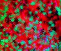
- Digital Images
- Online
Cellular architecture of normal human skin imaged by whole mount tissue microscopy. Human skin has a rich network of white blood cells (specifically dendritic cells, T cells and macrophages) which form sheaths around blood vessels. In this image, T cells (stained for CD3; red) dendritic cells (stained for MHC class II; green) and macrophages (stained for LYVE-1; blue with some cells showing a tinge of green) can be seen. Cell nuclei have been stained with DAPI (grey). This normal cellular architecture is grossly disrupted in diseased skin (see related images). X10 magnification. Scale bar (white) represents 200 micrometres.
Dr. Xiao-nong Wang, Human Dendritic Cell Laboratory, Newcastle University
- Digital Images
- Online
Cellular architecture of normal human skin imaged by whole mount tissue microscopy. Human skin has a rich network of white blood cells (specifically dendritic cells, T cells and macrophages) which form sheaths around blood vessels. In this image, T cells (stained for CD3; red) dendritic cells (stained for MHC class II; green) and macrophages (stained for LYVE-1; blue with some cells showing a tinge of green) can be seen. Cell nuclei have been stained with DAPI (grey). This normal cellular architecture is grossly disrupted in diseased skin (see related images). X20 magnification. Scale bar (white) represents 100 micrometres.
Dr. Xiao-nong Wang, Human Dendritic Cell Laboratory, Newcastle University
- Digital Images
- Online
Cellular architecture of normal human skin imaged by whole mount tissue microscopy. Human skin has a rich network of white blood cells (specifically dendritic cells, T cells and macrophages) which form sheaths around blood vessels. In this image, blood vessels (string-like structures stained for CD31; red), lymphatic vessels (ribbon-like structures stained for LYVE-1; blue) and dendritic cells (stained for CD11c; green) can be seen. Macrophages (stained for LYVE-1; blue) are also present. This normal cellular architecture is grossly disrupted in diseased skin (see related images). X10 magnification. Scale bar (white) represents 200 micrometres.
Dr. Xiao-nong Wang, Human Dendritic Cell Laboratory, Newcastle University
- Digital Images
- Online
Cellular architecture of normal human skin imaged by whole mount tissue microscopy. Human skin has a rich network of white blood cells (specifically dendritic cells, T cells and macrophages) which form sheaths around blood vessels. In this image, blood vessels (string-like structures stained for CD31; green), lymphatic vessels (ribbon-like structures stained for LYVE-1; blue) and T cells (stained for CD3; red) can be seen. T cells are only found around dermal blood vessels. Macrophages (stained for LYVE-1; blue) are also present. This normal cellular architecture is grossly disrupted in diseased skin (see related images). X10 magnification. Scale bar (white) represents 200 micrometres.
Dr. Xiao-nong Wang, Human Dendritic Cell Laboratory, Newcastle University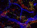
- Digital Images
- Online
Cellular architecture of normal human skin imaged by whole mount tissue microscopy. Human skin has a rich network of white blood cells (specifically dendritic cells, T cells and macrophages) which form sheaths around blood vessels (string-like structures). A network of lymphatic vessels (ribbon-like structures) is also present. In this image, human skin lymphatic vessels (stained for LYVE-1; blue) and white blood cells comprised of dendritic cells (stained for CD11c; green) and T cells (stained for CD3; red) can be seen. Some macrophages also express the protein LYVE-1 similar to lymphatic vessel cells which can be appreciated as blue cells within and in between the sheaths of white blood cells. This normal cellular architecture is grossly disrupted in diseased skin (see related images). X10 magnification. Scale bar (white) represents 200 micrometres.
Dr. Xiao-nong Wang, Human Dendritic Cell Laboratory, Newcastle University