Wellcome uses cookies.
Read our policy
Close cookie notification
Skip to main content
Wellcome Collection homepage
Visit us
What’s on
Stories
Collections
Get involved
About us
Sign in to your library account
Search our stories, images, catalogue and events
Library account
Search our stories, images, catalogue and events
Search
Images search
Search for images
Search
All
Stories
Images
Catalogue
Events
Colours
Licences
Creative Commons CC-BY (224)
Creative Commons CC-BY-NC (48)
Creative Commons CC0 (38)
Creative Commons CC-BY-NC-ND (1)
Public Domain Mark (1)
Dates
From
to
Types/Techniques
Etchings (1)
Subjects
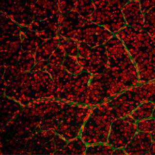
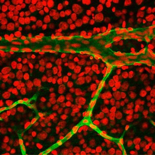
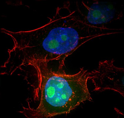
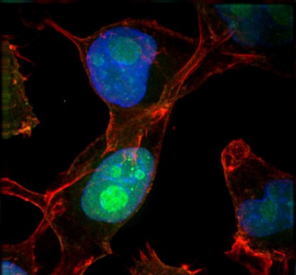
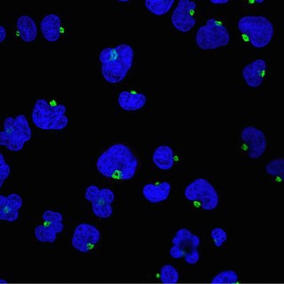
Nuclei (59)
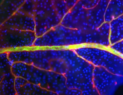
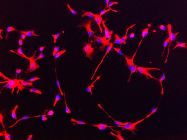
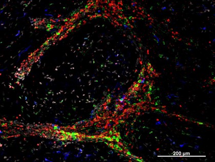
Blue (37)
Cancer (32)
Red (28)
Green (27)
Intestine (26)
Tumour (26)
Villi (26)
Nucleus (24)
Actin (20)
CONFOCAL (20)
DEVELOPMENT (20)
Malignancy (20)
Tumor (20)
Diarrhoea (19)
Pathogen (17)
Infectious disease (16)
Yellow (16)
E. coli (15)
IMMUNOFLUORESCENCE (15)
Contributors
S. Schuller (46)
Kevin Mackenzie, University of Aberdeen (17)
Rob Young (15)
Alex Gray (13)
Paul Appleton, University of Dundee (13)
Huw Parry & Michael Whitaker (8)
Lorna McInroy (8)
Alan Handyside (7)
Dr Jeremy Skepper (7)
NIMR, Francis Crick Institute (7)
University of Edinburgh (7)
Dr. S. Srinivas, Uni of Oxford (6)
Izzat Suffian, Pedro Costa, Stephen Pollard, David McCarthy & Khuloud T. Al-Jamal (6)
Royal Veterinary College (6)
A. Walker, L. Sharp & J. Pryde (4)
Anne Clark, University of Oxford (4)
S. Roy & C. Wolff (4)
Wellcome Images (4)
William R. Geddie (4)
Matthew Daniels (3)
Submit
312 results
Search result sorting
Sort by:
Relevance
Production dates
Sort order:
Ascending
Descending
Submit
Page
1
of 11
Next (page 2)
Close modal window
Page
1
of 11
Next (page 2)