Wellcome uses cookies.
Read our policy
Close cookie notification
Skip to main content
Wellcome Collection homepage
Visit us
What’s on
Stories
Collections
Get involved
About us
Sign in to your library account
Search our stories, images, catalogue and events
Library account
Search our stories, images, catalogue and events
Search
Images search
Search for images
Search
All
Stories
Images
Catalogue
Events
Colours
Licences
Creative Commons CC-BY (82)
Creative Commons CC-BY-NC (23)
Creative Commons CC0 (6)
In copyright (5)
Public Domain Mark (4)
Dates
From
to
Types/Techniques
Lithographs (4)
Acrylic paintings (2)
Caricatures (2)
Ephemera (2)
Intaglio prints (2)
Leaflets (2)
Posters (2)
Photographic prints (1)
Subjects
Model organism (53)
Aquatic vertebrae (24)
CNS (22)
Fruit fly (20)
Green (20)
Insect (16)
Pink (16)
Development (15)
GFP (14)
Magenta (14)
MODEL ORGANISM (14)
Purple (14)
Transgenic (14)
Neural network (13)
Neuronal signalling (11)
DEVELOPMENT (10)
Red (10)
Cyan (9)
Neuroscience (9)
Embryo (8)
Contributors
Fernan Federici & Jim Haseloff (16)
Derric Nimmo & Paul Eggleston (14)
Kate Turner, Dr Steve Wilson (12)
Anne Weston, Francis Crick Institute (10)
Abigail Tucker (9)
Ana Faro, Dr Steve Wilson (5)
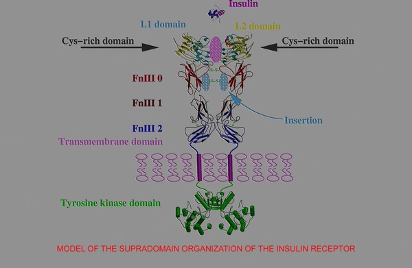
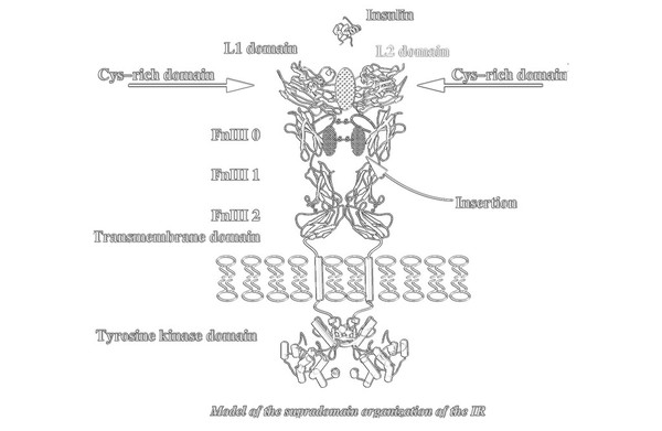
T.Blundell & N Campillo (5)
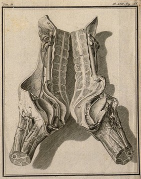
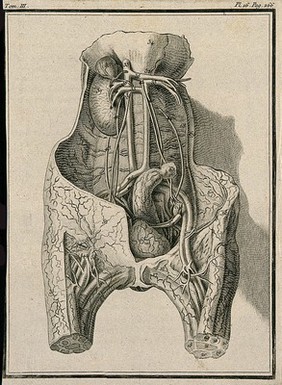
Buffon, Georges Louis Leclerc, comte de, 1707-1788 (4)
David Strutt (4)
Odra Noel (4)
Leo Valdivia, Dr Steve Wilson (3)
Scott Birch (3)
Alken, Henry Thomas, 1784-1851 (2)
Bump, L (2)
Combe, George, 1788-1858 (2)
Ennis, Richard, 1958- (2)
Macroscopic Solutions (2)
S. Roy & F. Muller (2)
S. Roy & S Higashijima (2)
Sawyer, H (2)
Submit
126 results
Search result sorting
Sort by:
Relevance
Production dates
Sort order:
Ascending
Descending
Submit
Page
1
of 5
Next (page 2)
Close modal window
Page
1
of 5
Next (page 2)