Wellcome uses cookies.
Read our policy
Close cookie notification
Skip to main content
Wellcome Collection homepage
Visit us
What’s on
Stories
Collections
Get involved
About us
Sign in to your library account
Search our stories, images, catalogue and events
Library account
Search our stories, images, catalogue and events
Search
Images search
Search for images
Search
All
Stories
Images
Catalogue
Events
Colours
Licences
Creative Commons CC-BY (27)
Creative Commons CC-BY-NC (18)
Public Domain Mark (6)
In copyright (1)
Dates
From
to
Types/Techniques
Engravings (3)
Book (1)
Photogravures (1)
Portrait prints (1)
Prints (1)
Subjects
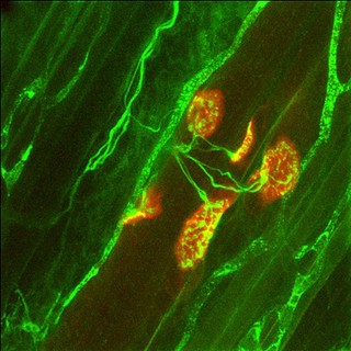
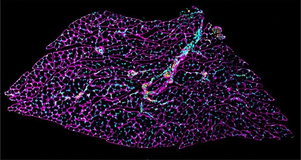
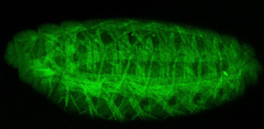
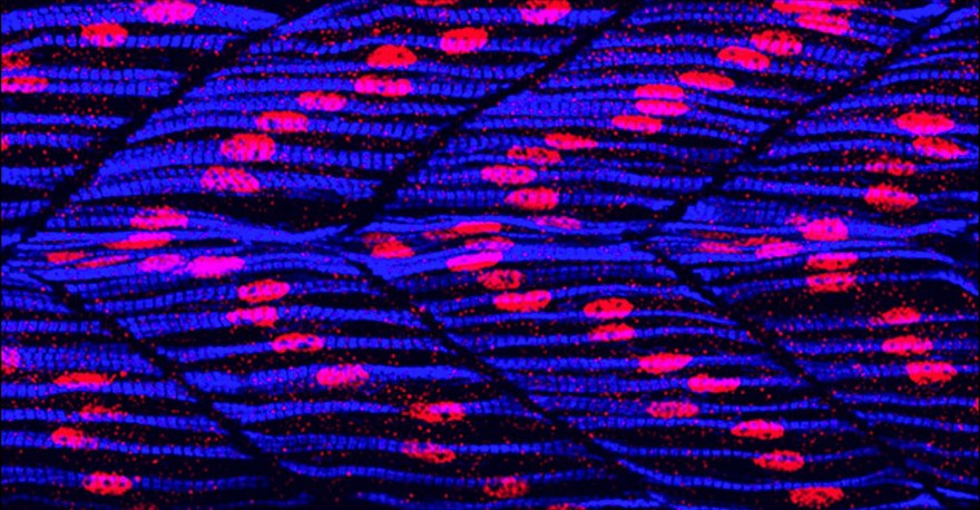
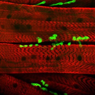
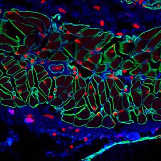
Muscle (13)
Nerves (9)
Structure (9)
Artwork (8)
Clay (8)
Cranium (8)
Forensic science (8)
Feel (7)
Neuron (7)
CONTRACTION (6)
Nerve (6)
Blue (5)
Bungarotoxin (5)
Extensor (5)
Extensor digitorum (5)
Fast-twitch muscle (5)
Fluorescent probe (5)
Mammal (5)
Rat (5)
Receptor (5)
Contributors
Heather Spears, photography ICandy (8)
Dr Guy Bewick, Aberdeen Univ (5)
Hermann Aberle, University of Munster (4)
Odra Noel (4)
S. Roy & C. Wolff (4)
Mike Kayser (3)
University of Edinburgh (3)
Blane, Gilbert, Sir, 1749-1834 (2)
Monro, Alexander, 1733-1817 (2)
Prof Giorgio Gabella (2)
Prof. Peter Brophy (2)
Bartholin, Thomas, 1616-1680 (1)
Bénard, 1731-1794 (1)
Bidloo, Govard, 1649-1713 (1)
Delamotte, William Alfred (1)
Diderot, Denis, 1713-1784 (1)
Dr Hermann Aberle, University of Munster (1)
Dr Jonathan Clarke (1)
Kevin Mackenzie, University of Aberdeen (1)
Laurence Jackson, Centre for Advanced Biomedical Imaging (1)
Submit
54 results
Search result sorting
Sort by:
Relevance
Production dates
Sort order:
Ascending
Descending
Submit
Page
1
of 2
Next (page 2)
Close modal window
Page
1
of 2
Next (page 2)


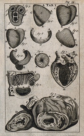


![Observations on the muscles, and particularly on the effects of their oblique fibres: with an appendix, in which the pretension of Dr. Gilbert Blane, that he first demonstrated the same effect to be produced by oblique muscles as by straight ones, with a less proportional decurtation of fibres, is proved to be unfounded ... / [Alexander Monro].](https://iiif.wellcomecollection.org/image/L0068866/full/282%2C/0/default.jpg)
![Observations on the muscles, and particularly on the effects of their oblique fibres: with an appendix, in which the pretension of Dr. Gilbert Blane, that he first demonstrated the same effect to be produced by oblique muscles as by straight ones, with a less proportional decurtation of fibres, is proved to be unfounded ... / [Alexander Monro].](https://iiif.wellcomecollection.org/image/L0068867/full/282%2C/0/default.jpg)