Wellcome uses cookies.
Read our policy
Close cookie notification
Skip to main content
Wellcome Collection homepage
Visit us
What’s on
Stories
Collections
Get involved
About us
Sign in to your library account
Search our stories, images, catalogue and events
Library account
Search our stories, images, catalogue and events
Search
Images search
Search for images
Search
All
Stories
Images
Catalogue
Events
Colours
Licences
Public Domain Mark (154)
In copyright (32)
Creative Commons CC-BY-NC (11)
Creative Commons CC-BY (6)
Dates
From
to
Types/Techniques
Photographs (133)
Albumen prints (78)
Gelatin silver prints (53)
Watercolors (18)
Drawings (13)
Prints (12)
Photomechanical prints (10)
Lithographs (9)
Paintings (7)
Engravings (5)
Chalk drawings (4)
Graphite drawings (4)
Portrait prints (4)
Etchings (3)
Group portraits (3)
Oil paintings (3)
Pencil works (3)
Portrait photographs (3)
Posters (3)
Cityscape photographs (2)
Subjects
Down syndrome (27)
St. Nicholas' and St. Martin's Orthopaedic Hospital (Surrey, England) (26)
Poetry (17)
Alchemy (16)
Astrology, Tibetan (12)
Mañjūśrī (12)
Predictive astrology (12)
Pechili Province (China) (11)
Beijing (China) (9)
Education (8)
London Institution (8)
Children with mental disabilities (5)
Typhoid fever (5)
Gas light fixtures (4)
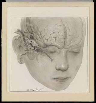
Human anatomy (4)
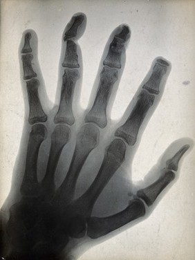
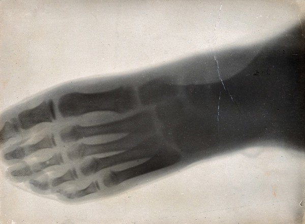
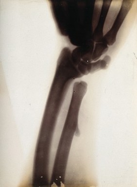
Human skeleton (4)
Leg (4)
London (England) (4)
Muscles (4)
Albert, Prince Consort, consort of Victoria, Queen of Great Britain, 1819-1861 (3)
Contributors
Shuttleworth, G. E. (George Edward), 1842-1928 (73)
Schuster, Arthur, Sir, 1851-1934 (32)
Thomson, J. (John), 1837-1921 (12)
Brooks, William, 1786-1867 (6)
Davis, John (Portrait and landscape photographer) (6)
Schnebbelie, Robert Blemmel, -approximately 1849 (5)
Michelangelo Buonarroti, 1475-1564 (4)
Armbruster, Johann Michael, 1761-1814 (3)
Brill, Reginald, 1902-1974 (3)
Gessner, Georg, 1765-1843 (3)
Holcroft, Thomas, 1745-1809 (3)
Lavater, Johann Caspar, 1741-1801 (3)
Lock & Whitfield (3)
May, N. Goulton (3)
Oakley (3)
Uganda Sleeping Sickness Commission. Meeting 1902) (3)
Walton, W. L., active 1834-1855 (3)
Bernardino, de Sahagún, 1499-1590 (2)
Royal Library of Berlin (2)
Submit
228 results
Search result sorting
Sort by:
Relevance
Production dates
Sort order:
Ascending
Descending
Submit
Page
1
of 8
Next (page 2)
Close modal window
Page
1
of 8
Next (page 2)