Wellcome uses cookies.
Read our policy
Close cookie notification
Skip to main content
Wellcome Collection homepage
Visit us
What’s on
Stories
Collections
Get involved
About us
Sign in to your library account
Search our stories, images, catalogue and events
Library account
Search our stories, images, catalogue and events
Search
Images search
Search for images
Search
All
Stories
Images
Catalogue
Events
Colours
Licences
Creative Commons CC-BY-NC (28)
Public Domain Mark (11)
Creative Commons CC-BY (5)
Creative Commons CC0 (1)
Dates
From
to
Types/Techniques
Banners - Tibet (5)
Distemper painting (5)
Engravings (5)
Paintings (5)
Tankas (Tibetan scrolls) (5)
Allegorical prints (2)
Book illustrations (2)
Lithographs (2)
Maps (2)
Caricatures (1)
Etchings (1)
Pencil works (1)
Subjects
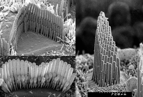
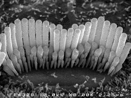
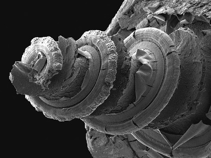
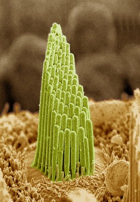
GUINEA PIG (10)
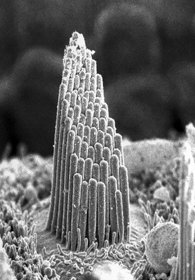
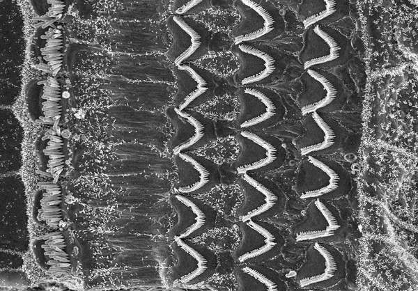
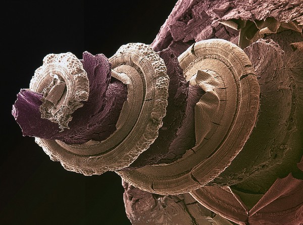
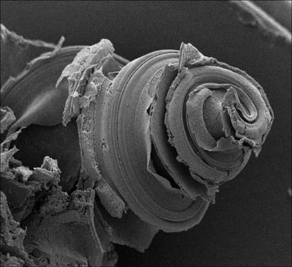
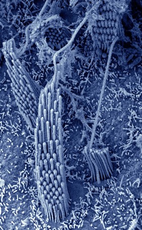
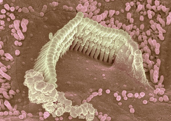
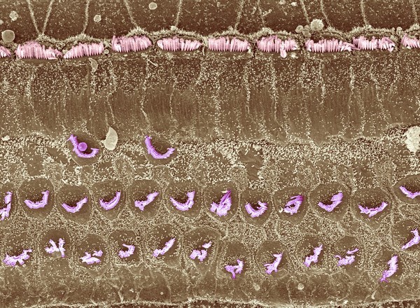
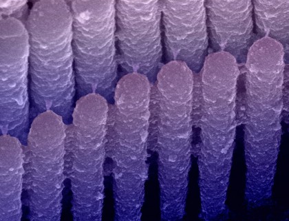
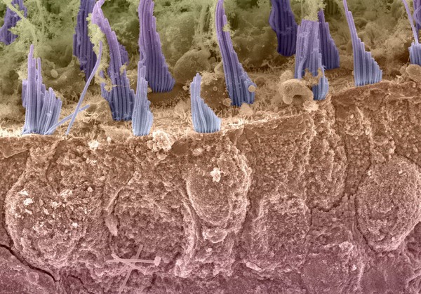
HAIR CELL (10)
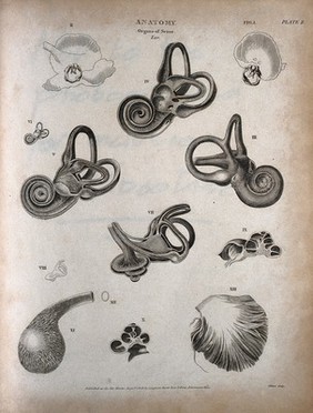
Inner ear (10)
AUDITORY (9)
Hair cells (8)
INNER EAR (8)
SCANNING ELECTRON MICROSCOPE (7)
Senses (7)
SENSORY HAIR BUNDLE (7)
BIOMEDICAL IMAGE AWARD (6)
Balance (5)
Buddhist gods (5)
SCANNING ELECTRON MICROGRAPH (5)
SEM (5)
TERRAPIN (5)
Human anatomy (4)
Stereocilia (4)
Vestibular organ (4)
Mechanoreceptor (3)
Side effects (3)
Contributors
Dr David Furness (25)
Caroline Gunn (2)
Hoskins, John, 1566-1638 (2)
Milton, Thomas, 1743?-1827 (2)
Akshay Kumar, Tom Davies and Nobue Itasaki, University of Bristol (1)
Buchanan, Thomas, 1782-1853 (1)
Consitt & Son (1)
De Wilde, Samuel, 1751-1832 (1)
Kirtland, George, active 1796-1815 (1)
Prof. Andrew Forge (1)
Steph Hares, University of Bristol (1)
Stewart, James (1)
Submit
50 results
Search result sorting
Sort by:
Relevance
Production dates
Sort order:
Ascending
Descending
Submit
Page
1
of 2
Next (page 2)
Close modal window
Page
1
of 2
Next (page 2)