Wellcome uses cookies.
Read our policy
Close cookie notification
Skip to main content
Wellcome Collection homepage
Visit us
What’s on
Stories
Collections
Get involved
About us
Sign in to your library account
Search our stories, images, catalogue and events
Library account
Search our stories, images, catalogue and events
Search
Images search
Search for images
Search
All
Stories
Images
Catalogue
Events
Colours
Licences
Public Domain Mark (14)
Creative Commons CC-BY (5)
Creative Commons CC0 (4)
In copyright (1)
Dates
From
to
Types/Techniques
Gelatin silver prints (4)
Photographs (4)
Lithographs (2)
Drawings (1)
Engravings (1)
Etchings (1)
Line photoengravings (1)
Photographic prints (1)
Subjects
19th Century (4)
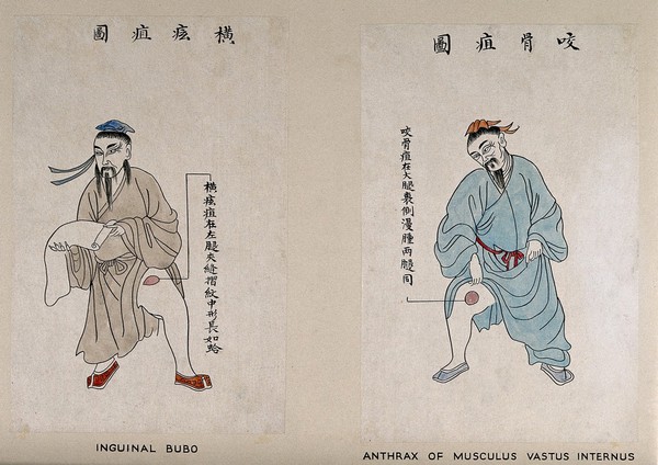
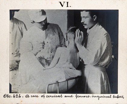
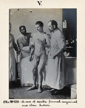
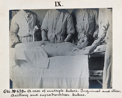
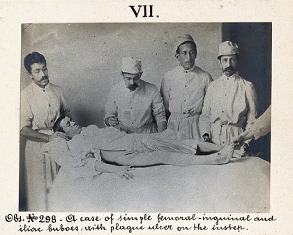
Plague (4)
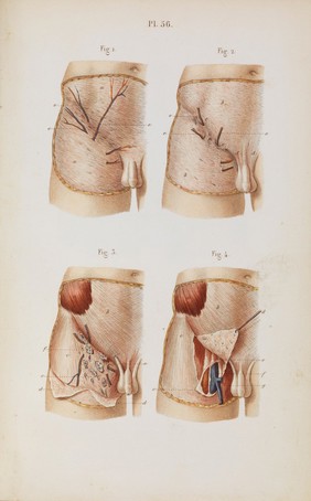
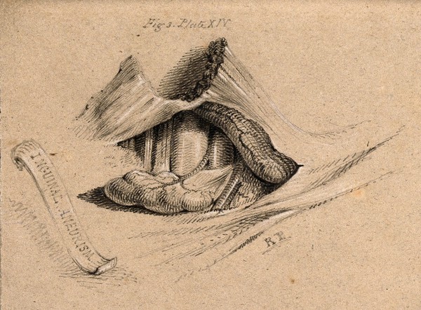
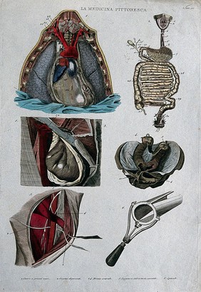
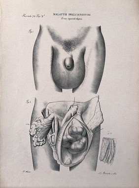
Surgery (4)
Antibiotics (3)
Brown (3)
Corkscrew (3)
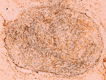
Dieterle stain (3)
Grey (3)
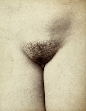
Human anatomy (3)
Inguinal (3)
Orange (3)
Pattern (3)
Sample (3)
Section (3)
Spiral (3)
Spirochaete (3)
Spirochete (3)
STI (3)
Wavy (3)
Anatomy (2)
Contributors
St Bartholomew's Hospital Archives & Museum (4)
William R. Geddie (3)
Batelli (1)
Delamotte, William Alfred (1)
Dr Henry Oakeley (1)
Godart, Thomas (1)
Maclise, Joseph (1)
Méheux, Félix (1)
Mitchell, E., active approximately 1811 (1)
Muzzi, Ottavio (1)
SB Lucas (1)
Submit
24 results
Search result sorting
Sort by:
Relevance
Production dates
Sort order:
Ascending
Descending
Submit
Page
1
of 1
Close modal window
Page
1
of 1