Wellcome uses cookies.
Read our policy
Close cookie notification
Skip to main content
Wellcome Collection homepage
Visit us
What’s on
Stories
Collections
Get involved
About us
Sign in to your library account
Search our stories, images, catalogue and events
Library account
Search our stories, images, catalogue and events
Search
Images search
Search for images
Search
All
Stories
Images
Catalogue
Events
Colours
Licences
Public Domain Mark (19)
Dates
From
to
Types/Techniques
Diagrams (15)
Ink drawings (12)
Drawings (8)
Paintings (8)
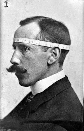
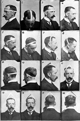
Photographs (4)
Photomechanical prints (4)
Prints (4)
Lithographs (2)
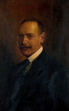
Oil paintings (2)
Portrait paintings (2)
Engravings (1)
Pencil works (1)
Portrait drawings (1)
Watercolors (1)
Subjects
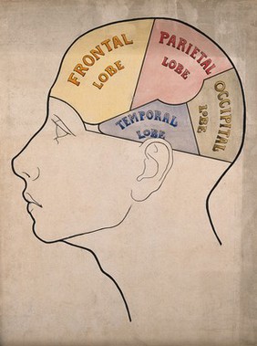
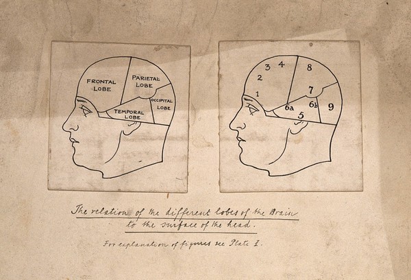
Phrenology (22)
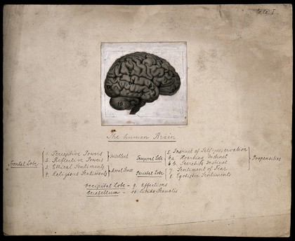
Brain (20)
Psychology - History (20)
Neurophysiology (19)
Head (17)
Brain - Localisation of functions (15)
Craniology (11)
Skull (9)
Physiognomy (7)
Characters and characteristics (6)
Hollander, Bernard, 1864-1934 (6)
Brain - Anatomy (4)
Cerebellum (4)
Children - Diseases (4)
Craniometry (4)
Measurement (4)
Measuring instruments (4)
Intellect (3)
People with mental disabilities (3)
Sex differences (3)
Contributors
Hollander, Bernard, 1864-1934 (20)
Engelmann, G. (Godefroy), 1788-1839 (2)
Gall, F. J. (Franz Joseph), 1758-1828 (2)
Mazer, Carl Peter, 1807-1884 (2)
Vimont, Joseph, 1795-1857 (2)
Caldwall, James, 1739-1819 (1)
Reid, Robert W. (Robert William), 1851-1939 (1)
Webber, John, 1751-1793 (1)
Submit
25 results
Search result sorting
Sort by:
Relevance
Production dates
Sort order:
Ascending
Descending
Submit
Page
1
of 1
Close modal window
Page
1
of 1