Wellcome uses cookies.
Read our policy
Close cookie notification
Skip to main content
Wellcome Collection homepage
Visit us
What’s on
Stories
Collections
Get involved
About us
Sign in to your library account
Search our stories, images, catalogue and events
Library account
Search our stories, images, catalogue and events
Search
Images search
Search for images
Search
All
Stories
Images
Catalogue
Events
Colours
Licences
Creative Commons CC-BY (128)
Public Domain Mark (66)
In copyright (33)
Creative Commons CC-BY-NC (25)
Creative Commons CC0 (18)
Dates
From
to
Types/Techniques
Ephemera (27)
Engravings (20)
Leaflets (12)
Blotting paper (11)
Broadsides (7)
Calendars (6)
Case studies (5)
Lithographs (5)
Photographs (5)
Portrait photographs (5)
Watercolors (5)
Book illustrations (4)
Photographic prints (4)
Paintings (3)
Prints (3)
Direct mail (2)
Drawings (2)
Gelatin silver prints (2)
Intaglio prints (2)
Manufacturers' catalogues (2)
Subjects
Red (28)
Reproductive system (23)
Artwork (22)
Concept (22)
Abstract (21)
Bleeding (21)
Blood flow (21)
Cycles (21)
Female (21)
Float (21)
Fluid (21)
Gynaecology (21)
Period pain (21)
Phase (21)
Sexual Health (21)
Human anatomy (18)
Drug Industry (16)
Pharmaceutical Preparations (16)
Gland (15)
Product (15)
Contributors
St Bartholomew's Hospital Archives & Museum (50)
Beauty in Blood (21)
Godart, Thomas (19)
Royal Veterinary College (8)
Mark, Leonard Portal (7)
SB Lucas (7)
Blankaart, Steven, 1650-1702 (6)
Burroughs Wellcome Company (6)
Caroline Gunn (6)
Fairchild Bros. & Foster (6)
S. Schuller (6)
Addison, Thomas, 1793-1860 (5)
Hall, William Henry, -1807 (5)
Allen & Hanburys (4)
Delamotte, William Alfred (4)
Endo Products, Inc (4)
Ethical Products (4)
St Bartholomew's Hospital Photographic Society (4)
Bidloo, Govard, 1649-1713 (3)
University of Edinburgh (3)
Submit
288 results
Search result sorting
Sort by:
Relevance
Production dates
Sort order:
Ascending
Descending
Submit
Page
1
of 10
Next (page 2)
Close modal window
Page
1
of 10
Next (page 2)


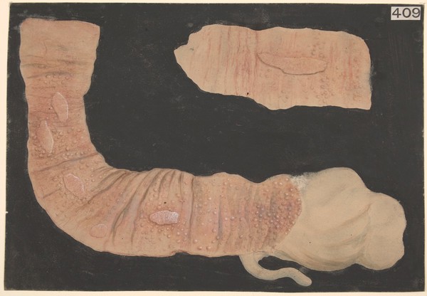


![Drawing of the 1918 Influenza: Trachea showing particular retention of epithelium, by, and in the neighbourhood of duet [?] of one of the mucus glands.](https://iiif.wellcomecollection.org/image/L0041258/full/282%2C/0/default.jpg)

