Wellcome uses cookies.
Read our policy
Close cookie notification
Skip to main content
Wellcome Collection homepage
Visit us
What’s on
Stories
Collections
Get involved
About us
Sign in to your library account
Search our stories, images, catalogue and events
Library account
Search our stories, images, catalogue and events
Search
Images search
Search for images
Search
All
Stories
Images
Catalogue
Events
Colours
Licences
Public Domain Mark (52)
Creative Commons CC-BY (16)
In copyright (12)
Creative Commons CC-BY-NC (1)
Dates
From
to
Types/Techniques
Watercolors (40)
Paintings (34)
Drawings (26)
Acrylic paintings (10)
Advertisements (10)
Mural painting and decoration (10)
Lithographs (8)
Atlases (Scientific) (7)
Posters (7)
Chromolithographs (5)
Engravings (4)
Ephemera (4)
Charts (2)
Glamour photographs (2)
Photographic prints (2)
Photographs (2)
Electronic journals (1)
Portrait photographs (1)
Stipple engravings (1)
Subjects
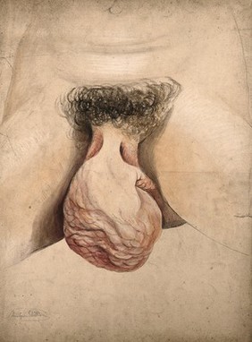
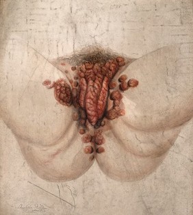
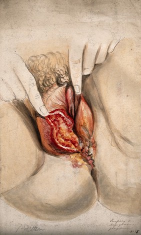
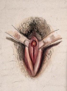
Royal Free Hospital (London, England) (38)
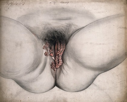
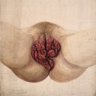
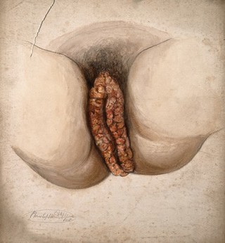
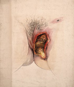
Genitalia, Female (12)
Benin (10)
Sexually transmitted diseases (10)
Vodou (10)
Women (10)
Genital Diseases, Female (7)
Gynecology (7)
Pathology, Surgical (7)
Female genital mutilation (5)
Human anatomy (4)
Pregnancy (4)
Uterus, Pregnant (4)
London (England) (3)
Sudan (3)
Abnormalities, Human (2)
Cuba (2)
Djibouti (2)
Drug Industry (2)
Endometritis - drug therapy (2)
Contributors
D'Alton, Christopher, active 1847-1871 (39)
Savage, Henry, -1900 (9)
Fallaize, Edwin Nichol, 1877-1957 (2)
Godart, Thomas (2)
Gri︠u︡n, O., active 1919 (2)
Hunter, William, 1718-1783 (2)
Laboratorios Vieta-Plasencia (2)
Liceti, Fortunio, 1577-1657. De monstris (2)
Liceti, Fortunio, 1577-1657. De monstris. French (2)
Lizars, W. H. (William Home), 1788-1859 (2)
Mauriceau, François, 1637-1709 (2)
Palfijn, Jan, 1650-1730 (2)
Palfijn, Jan, 1650-1730. Description anatomique de la disposition surprenante de quelques parties externes & internes de deux enfans (2)
Robinson & Sons Ltd. (Chesterfield, England) (2)
Soviet Union. Otdel okhrany materinstva i mladenchestva (2)
St Bartholomew's Hospital Archives & Museum (2)
Blakey (1)
Hana Studios Ltd (1)
Moreau-Valvile, active 1806 (1)
Tresca, Salvadore, 1750?-1815 (1)
Submit
92 results
Search result sorting
Sort by:
Relevance
Production dates
Sort order:
Ascending
Descending
Submit
Page
1
of 4
Next (page 2)
Close modal window
Page
1
of 4
Next (page 2)