Skip to main content
Wellcome Collection homepage
Visit us
What’s on
Stories
Collections
Get involved
About us
Sign in to your library account
Search our stories, images, catalogue and events
Library account
Search our stories, images, catalogue and events
Search
Images search
Search for images
Search
All
Stories
Images
Catalogue
Events
Colours
Licences
Creative Commons CC-BY (91)
Creative Commons CC-BY-NC (16)
Creative Commons CC0 (1)
Dates
From
to
Types/Techniques
Subjects
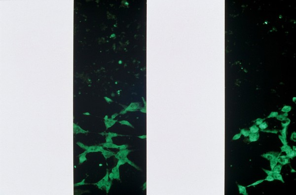
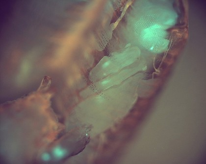
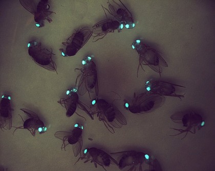
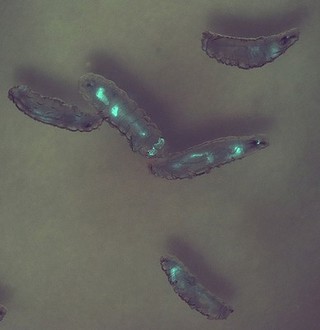
GFP (33)
Model organism (21)
Transgenic (18)
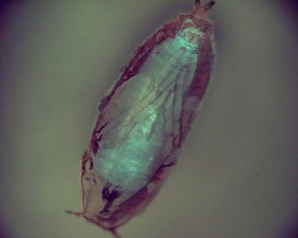
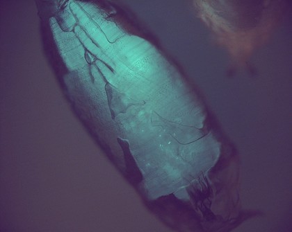
Fruit fly (17)
Insect (16)
Green fluorescent protein (13)
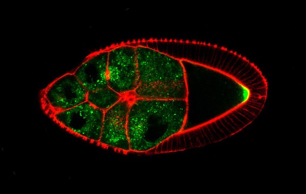
Development (11)
Embryo (11)
Green (10)
DAPI (8)
IMMUNOFLUORESCENCE (8)
Red (8)
Muscle (7)
Blue (6)
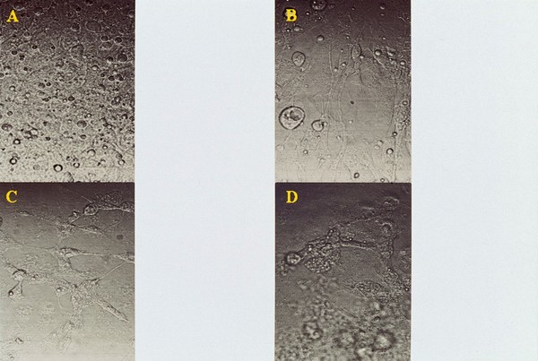
CANCER CELLS (5)
Metamorphosis (5)
Microbe (5)
Morphology (5)
Pattern (5)
Shape (5)
Contributors
Fernan Federici & Jim Haseloff (23)
Derric Nimmo & Paul Eggleston (17)
Alex Gray (8)
Biosciences Imaging Gp, Soton (4)
Fernán Federici (4)
Hermann Aberle, University of Munster (4)
Kate Turner (4)
Kate Turner, Dr Steve Wilson (3)
Monica Folgueira & Steve Wilson (3)
Monica Folguiera (3)
Alison Dun, ESRIC (Edinburgh Super-Resolution Imaging Consortium) (2)
Dr Mónica Folgueira (2)
Dr Steve Wilson (2)
Dr. S. Srinivas, Uni of Oxford (2)
Kate Storey (2)
S. Roy & F. Muller (2)
S. Roy & S Higashijima (2)
Dr Andrea Brand and Boris Egger (1)
Torre-Ubieta, Luis de la (1)
Yirui Sun (1)
Submit
108 results
Search result sorting
Sort by:
Relevance
Production dates
Sort order:
Ascending
Descending
Submit
Page
1
of 4
Next (page 2)
Close modal window
Page
1
of 4
Next (page 2)