Wellcome uses cookies.
Read our policy
Close cookie notification
Skip to main content
Wellcome Collection homepage
Visit us
What’s on
Stories
Collections
Get involved
About us
Sign in to your library account
Search our stories, images, catalogue and events
Library account
Search our stories, images, catalogue and events
Search
Images search
Search for images
Search
All
Stories
Images
Catalogue
Events
Colours
Licences
Creative Commons CC-BY (17)
Creative Commons CC-BY-NC (3)
Creative Commons CC0 (2)
Dates
From
to
Types/Techniques
Subjects
Green (13)
Aquatic vertebrae (11)
Cyan (11)
Model organism (11)
CNS (10)
Magenta (10)
Purple (10)
Pink (9)
Neural network (8)
Neuronal signalling (5)
Blue (4)
Development (4)
Forebrain (4)
Neuroscience (4)
Bilateral (3)
Left-right brain asymmetry (3)
Mouse (3)
Red (3)
Behaviour (2)
MOLECULAR MODEL (2)
Contributors
Kate Turner, Dr Steve Wilson (6)
Ana Faro, Dr Steve Wilson (2)
Daniela Malide, Jean-Yves Metais, Cynthia E Dunbar, NIH, Bethesda, USA (2)
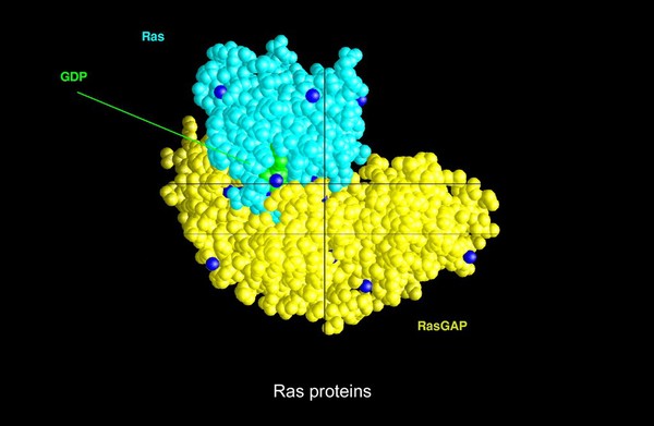
T.Blundell & N Campillo (2)
Ana Faro, Tom Hawkins and Dr Steve Wilson (1)
Ansel Oommen (1)
Anya Suppermpool, Rihel lab/ Wilson lab, University College London (1)
Dr Kara L.Cerveny & Dr Steve W.Wilson (1)
Ingrid Lekk, Dr Steve Wilson (1)
James N. Sleigh (1)
John Grady, Doug Turnbull, Claudia Racca, Newcastle University (1)
Kuan-Chung Su, London Research Institute, Cancer Research UK (1)
Lekk, Ingrid (1)
Mol. Biophysics, Oxford Univ (1)
Wellcome Images (1)
Wilson, Steve (1)
Submit
22 results
Search result sorting
Sort by:
Relevance
Production dates
Sort order:
Ascending
Descending
Submit
Page
1
of 1
Close modal window
Page
1
of 1