Wellcome uses cookies.
Read our policy
Close cookie notification
Skip to main content
Wellcome Collection homepage
Visit us
What’s on
Stories
Collections
Get involved
About us
Sign in to your library account
Search our stories, images, catalogue and events
Library account
Search our stories, images, catalogue and events
Search
Images search
Search for images
Search
All
Stories
Images
Catalogue
Events
Colours
Licences
Creative Commons CC-BY-NC (87)
Creative Commons CC-BY (76)
Public Domain Mark (25)
Creative Commons CC0 (15)
In copyright (15)
Dates
From
to
Types/Techniques
Book illustrations (16)
Lithographs (6)
Paintings (5)
Posters (5)
Watercolors (5)
Etchings (3)
Intaglio prints (3)
Aquatints (2)
Chromolithographs (2)
Drawings (2)
Engravings (2)
Periodical illustrations (2)
Broadsides (1)
Cards (1)
Ephemera (1)
Landscape prints (1)
Mezzotints (1)
Museum object (1)
Novelty works (1)
Soft-ground etchings (1)
Subjects
Clusters (67)
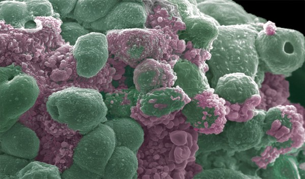
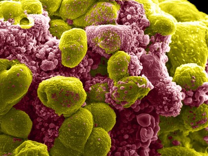
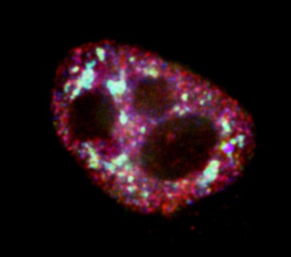
Cancer (48)
Tumor (41)
Malignancy (40)
Tumour (39)
Blue (38)
Spheres (34)
Green (32)
Ball (31)
Red (28)
Purple (24)
Immunology (22)
Pink (20)
Ruffles (20)
Yellow (19)
Development (18)
Blebbing (17)
Immortal (16)
Chinese Medicine (15)
TCM (15)
Contributors
Rowan McOnegal (30)
Anne Weston, Francis Crick Institute (26)
Annie Cavanagh (10)
Dr. Xiao-nong Wang, Human Dendritic Cell Laboratory, Newcastle University (9)
Prof. Andrew Forge (9)
Jenny Nichols (7)
John Grady, Doug Turnbull, Claudia Racca, Newcastle University (5)
Royal Veterinary College (4)
Sue Snell (4)
D'Alton, Christopher, active 1847-1871 (3)
David Gregory & Debbie Marshall (3)
Jacquin, Nikolaus Joseph, Freiherr von, 1727-1817 (3)
Khuloud T. Al-Jamal & Houmam Kafa (3)
Khuloud T. Al-Jamal, David McCarthy & Izzat Suffian (3)
Michael Frank, Royal Veterinary College (3)
Scheidl, Franz Anton von, 1731-1801 (3)
Valeria Molinari, Louise Howell, Maria Vinci, Katy Taylor and Chris Jones, Institute of Cancer Research (3)
Alan Handyside (2)
Karen Gustafson (2)
Khuloud T. Al-Jamal & Izzat Suffian (2)
Submit
219 results
Search result sorting
Sort by:
Relevance
Production dates
Sort order:
Ascending
Descending
Submit
Page
1
of 8
Next (page 2)
Close modal window
Page
1
of 8
Next (page 2)
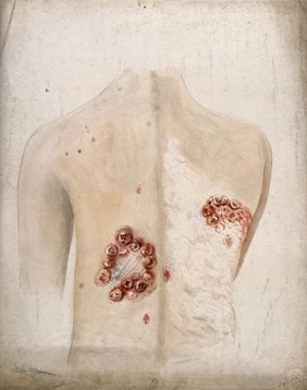


![Numerous hands clustered together with the words 'Wir brauchen Dich' [we need you] representing an advertisement for the centenary of the Berliner AIDS-Hilfe e.V. and an appeal for volunteers. Colour lithograph by Carolyn Jones and Comdesign, [1995].](https://iiif.wellcomecollection.org/image/b16742990_l0053614.jp2/full/600%2C/0/default.jpg)