Wellcome uses cookies.
Read our policy
Close cookie notification
Skip to main content
Wellcome Collection homepage
Visit us
What’s on
Stories
Collections
Get involved
About us
Sign in to your library account
Search our stories, images, catalogue and events
Library account
Search our stories, images, catalogue and events
Search
Images search
Search for images
Search
All
Stories
Images
Catalogue
Events
Colours
Licences
Public Domain Mark (21)
Creative Commons CC-BY (4)
In copyright (1)
Dates
From
to
Types/Techniques
Intaglio prints (5)
Engravings (4)
Pencil works (4)
Drawings (3)
Watercolors (3)
Gouaches (2)
Portrait paintings (2)
Wood engravings (2)
Book illustrations (1)
Collotypes (1)
Etchings (1)
Ink drawings (1)
Mezzotints (1)
Paintings (1)
Photographic prints (1)
Photographs (1)
Prints (1)
Radiographs (1)
Subjects
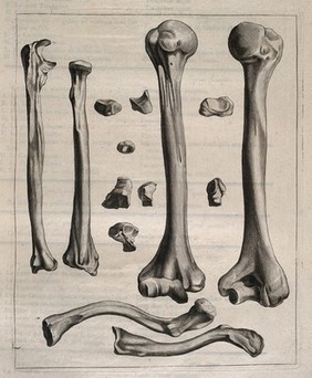
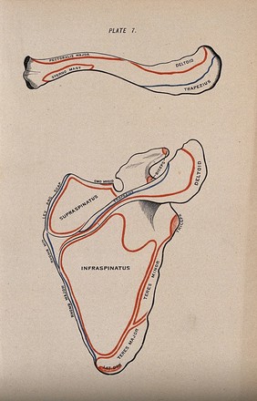
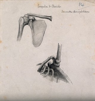
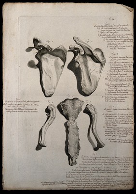
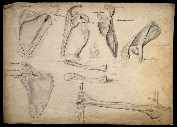
Human anatomy (11)
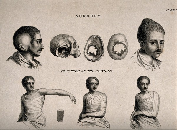
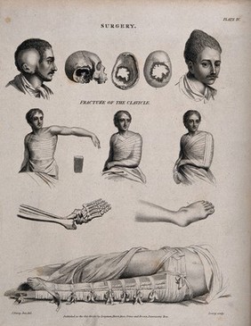
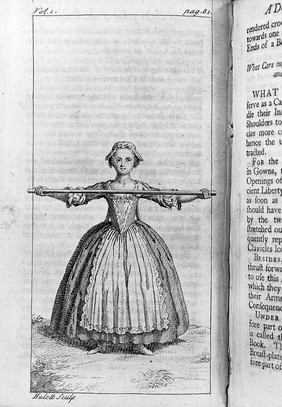
Clavicle (7)
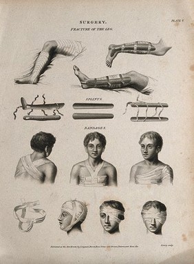
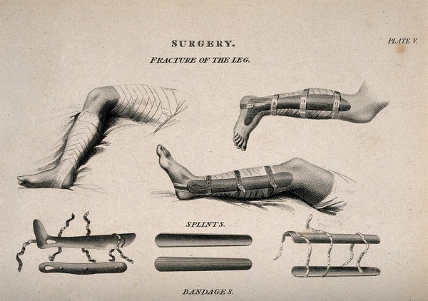
Bandages and bandaging (5)
Foot (5)
Fractures (5)
Leg (5)
Splints (Surgery) (5)
Wounds and injuries - Treatment (5)
Anatomy (4)
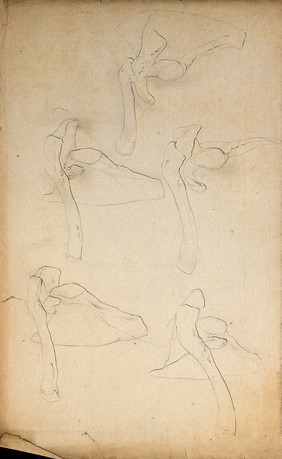
Bones (4)
Eye (3)
Head (3)
Human body (3)
Muscles (3)
19th Century (2)
Canton Hospital (Guangzhou, China) (2)
Dissection (2)
Head - Abnormalities (2)
Human skeleton (2)
Male (2)
Contributors
Farey, John, 1791-1851 (6)
Lowry, Wilson, 1762-1824 (6)
St Bartholomew's Hospital Archives & Museum (3)
Bidloo, Govard, 1649-1713 (2)
Cowper, William, 1666-1709 (2)
Godart, Thomas (2)
Lairesse, Gérard de, 1640-1711 (2)
Lam, Qua (2)
Cheselden, William, 1688-1752 (1)
Chinnery, E. W. Pearson (Ernest William Pearson), 1887-1972 (1)
D'Alton, Christopher, active 1847-1871 (1)
Duverney, M. (Jacques-François-Marie), 1661-1748 (1)
James Hulett (1)
Leisewitz, Theodor, active 1908, Assistant physician of the Royal Womens Hospital, Dresden (1)
Luo Shaoji (1)
Mongrédien, Adêle (1)
Römmler and Jonas (1)
Vesalius, Andreas, 1514-1564 (1)
Vesling, Johann, 1598-1649 (1)
Whishaw, J. C., 1833-1895 (1)
Submit
31 results
Search result sorting
Sort by:
Relevance
Production dates
Sort order:
Ascending
Descending
Submit
Page
1
of 2
Next (page 2)
Close modal window
Page
1
of 2
Next (page 2)