Skip to main content
Wellcome Collection homepage
Visit us
What’s on
Stories
Collections
Get involved
About us
Sign in to your library account
Search our stories, images, catalogue and events
Library account
Search our stories, images, catalogue and events
Search
Images search
Search for images
Search
All
Stories
Images
Catalogue
Events
Colours
Licences
Public Domain Mark (77)
Creative Commons CC-BY (58)
Creative Commons CC-BY-NC (28)
In copyright (12)
Creative Commons CC0 (6)
Dates
From
to
Types/Techniques
Book illustrations (40)
Engravings (9)
Lithographs (9)
Photographic prints (5)
Stereographs (5)
Watercolors (5)
Caricatures (4)
Drawings (4)
Posters (4)
Etchings (3)
Ink drawings (3)
Intaglio prints (3)
Paintings (3)
Soft-ground etchings (3)
Wood engravings (3)
Albumen prints (2)
Charts (2)
Ephemera (2)
Photographs (2)
Portrait prints (2)
Subjects
Channels (14)
Acu-moxa (11)
Ming period (1368-1644) (10)
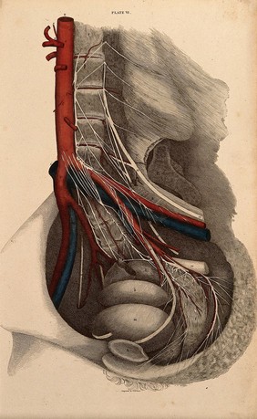
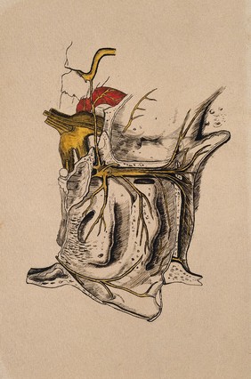
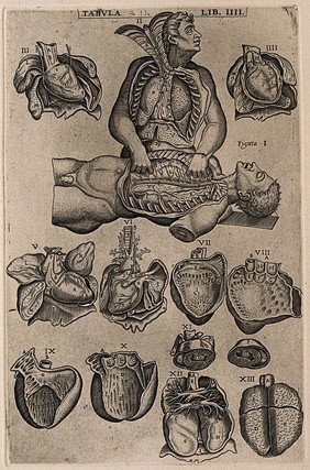
Human anatomy (9)
Brown (8)
Cavity (8)
Structure (8)
Hole (7)
Yellow (7)
Acupuncture chart (6)
CT scan (6)
Pain (6)
Cancer (5)
Tumour (5)
BINDING PROTEIN (4)
Devil (4)
Female (4)
MOLECULAR MODEL (4)
MOLECULE (4)
Neoplasm (4)
Contributors
Godart, Thomas (11)
St Bartholomew's Hospital Archives & Museum (11)
Justyna Miszkiewicz, Jayashree Chakraborty, John Logan, Duncan Bassett, Graham Williams, Imperial College London (5)
Waterston, D. (David) (5)
Peter Artymiuk (4)
Delamotte, William Alfred (3)
Gillray, James, 1756-1815 (3)
Macroscopic Solutions (3)
Michael Frank, Royal Veterinary College (3)
Royal Veterinary College (3)
Carole Reeves (2)
Clarke, Edward Daniel, 1769-1822 (2)
Cosway, Richard, 1742-1821 (2)
Luo Shaoji (2)
Midgley, Julia (2)
Orme, Daniel, 1766-1837 (2)
Schuster, Arthur, Sir, 1851-1934 (2)
Annie Cavanagh (1)
S. Roy (1)
Vesalius, Andreas, 1514-1564 (1)
Submit
185 results
Search result sorting
Sort by:
Relevance
Production dates
Sort order:
Ascending
Descending
Submit
Page
1
of 7
Next (page 2)
Close modal window
Page
1
of 7
Next (page 2)