Wellcome uses cookies.
Read our policy
Close cookie notification
Skip to main content
Wellcome Collection homepage
Visit us
What’s on
Stories
Collections
Get involved
About us
Sign in to your library account
Search our stories, images, catalogue and events
Library account
Search our stories, images, catalogue and events
Search
Images search
Search for images
Search
All
Stories
Images
Catalogue
Events
Colours
Licences
Creative Commons CC-BY (12)
Public Domain Mark (12)
Creative Commons CC-BY-NC (1)
Dates
From
to
Types/Techniques
Engravings (3)
Gouaches (1)
Ink drawings (1)
Lithographs (1)
Photographs (1)
Portrait paintings (1)
Portrait photographs (1)
Subjects
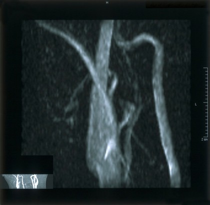
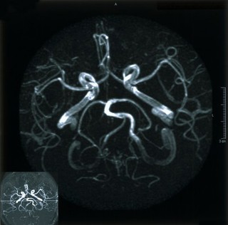
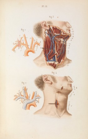
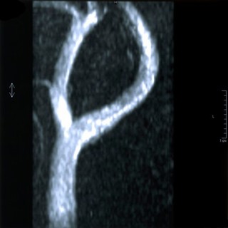
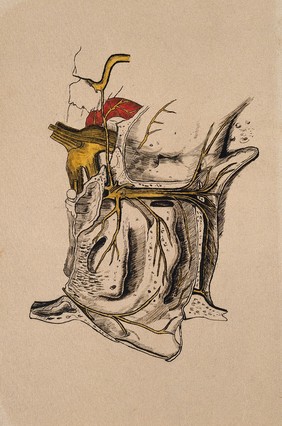
Human anatomy (4)
Male (4)
19th Century (3)
Dissection (3)
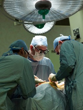
Surgery (3)
Anatomy (2)
Arteries (2)
Facemask (2)
Healthcare (2)
Healthcare professional (2)
Hospital (2)
Medical (2)
Operation (2)
Surgeon (2)
Theatre (2)
Veins (2)
Artery (1)
BALLOON ANGIOPLASTY (1)
Belladonna (Plant) (1)
BLOCKED ARTERY (1)
Contributors
Adrian Wressell, Heart of England NHS FT (2)
Bartholin, Thomas, 1616-1680 (1)
Blankaart, Steven, 1650-1702 (1)
Cane, A. A (1)
Delamotte, William Alfred (1)
Dr Andrew Loesch (1)
Evans, Nicholas (1)
Gray, Henry, 1827-1861 (1)
J. Basire (1)
Lam, Qua (1)
Lower, Richard, 1631-1691 (1)
Madeley, George Edward (1)
Michael Frank (1)
Michael Frank, Royal Veterinary College (1)
Nicholas Evans, University of Cambridge (1)
University of Cambridge (1)
Wellcome Images (1)
Willis, Thomas, 1621-1675 (1)
Submit
25 results
Search result sorting
Sort by:
Relevance
Production dates
Sort order:
Ascending
Descending
Submit
Page
1
of 1
Close modal window
Page
1
of 1