Wellcome uses cookies.
Read our policy
Close cookie notification
Skip to main content
Wellcome Collection homepage
Visit us
What’s on
Stories
Collections
Get involved
About us
Sign in to your library account
Search our stories, images, catalogue and events
Library account
Search our stories, images, catalogue and events
Search
Images search
Search for images
Search
All
Stories
Images
Catalogue
Events
Colours
Licences
Creative Commons CC-BY (52)
Creative Commons CC-BY-NC (51)
In copyright (7)
Creative Commons CC0 (3)
Creative Commons CC-BY-NC-ND (1)
Dates
From
to
Types/Techniques
Lithographs (7)
Posters (7)
Subjects
Projections (36)
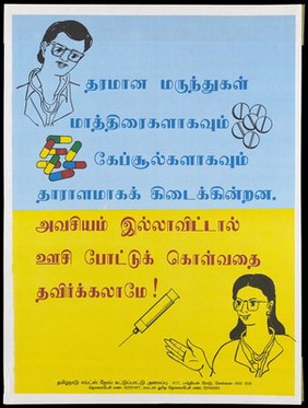
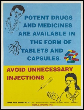
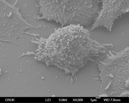
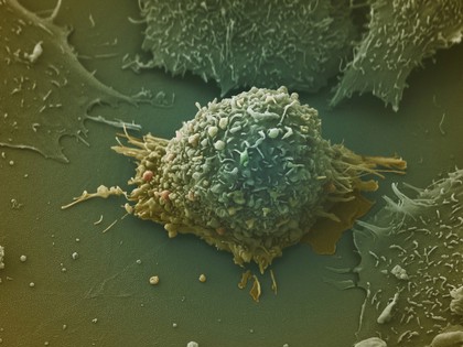
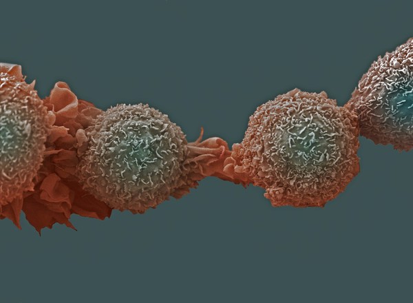
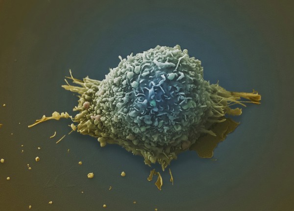
Cancer (35)
Blue (26)
Ruffles (26)
Cell culture (24)
Purple (19)
Green (18)
Malignancy (18)
Tumor (18)
Tumour (18)
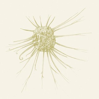
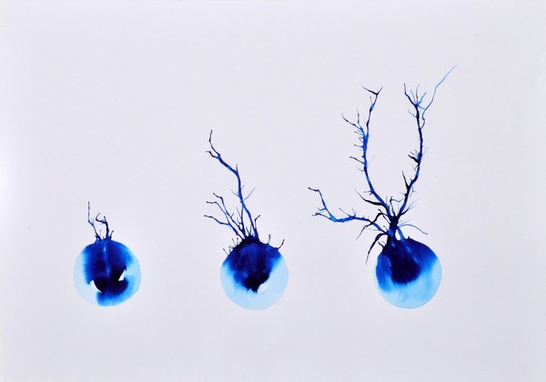
Cell (17)
Proliferation (17)
Development (15)
Environmental cancer (15)
Lung cancer (15)
Membrane processes (15)
Pulmonary (15)
Cell body (14)
Filopodia (14)
Immunology (14)
Contributors
Anne Weston, Francis Crick Institute (30)
Laura Trinkle-Mulcahy (10)
S. Schuller (7)
Izzat Suffian, Pedro Costa, Stephen Pollard, David McCarthy & Khuloud T. Al-Jamal (6)
David Goulding, Wellcome Trust Sanger Institute (5)
Mikaela Laine, University of Helsinki (4)
UNICEF (4)
Annie Cavanagh (3)
Judith Sleeman (3)
Stephen Magrath (3)
Yirui Sun (3)
Dr David Furness (2)
Dr Mónica Folgueira (2)
Errin Johnson (2)
Kevin Mackenzie, University of Aberdeen (2)
Monica Folguiera (2)
Rob Young (2)
Queen's University Belfast (1)
S. Roy (1)
University of Edinburgh (1)
Submit
114 results
Search result sorting
Sort by:
Relevance
Production dates
Sort order:
Ascending
Descending
Submit
Page
1
of 4
Next (page 2)
Close modal window
Page
1
of 4
Next (page 2)