Wellcome uses cookies.
Read our policy
Close cookie notification
Skip to main content
Wellcome Collection homepage
Visit us
What’s on
Stories
Collections
Get involved
About us
Sign in to your library account
Search our stories, images, catalogue and events
Library account
Search our stories, images, catalogue and events
Search
Images search
Search for images
Search
All
Stories
Images
Catalogue
Events
Colours
Licences
Creative Commons CC-BY (55)
Creative Commons CC-BY-NC (19)
Creative Commons CC0 (14)
Public Domain Mark (13)
In copyright (1)
Dates
From
to
Types/Techniques
Book illustrations (5)
Prints (5)
Collotypes (4)
Radiographs (4)
Lithographs (2)
Engravings (1)
Museum object (1)
Photomechanical prints (1)
Wood engravings (1)
Subjects
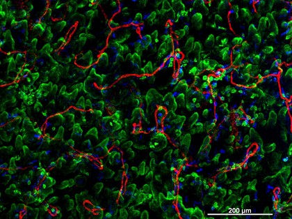
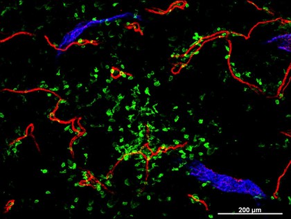
Red (26)
Structure (24)
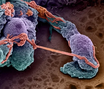
3D Imaging (14)
BriteVu (14)
Contrast agent (14)
CT scan (14)
Digital imaging (14)
Imaging (14)
Vascular system (14)
Vasculature (14)
Morphology (13)
Blue (11)
Orange (11)
Specimen (11)
White (11)
Animal (9)
Black (9)
Dye (9)
Thermoregulation (9)
Mouse (8)
Contributors
Scott Echols (11)
David Linstead (10)
Rob Young (8)
Izzat Suffian, Houmam Kafa, David McCarthy & Khuloud T. Al-Jamal (4)
Jean Wade and Linda Sharp (4)
Leisewitz, Theodor, active 1908, Assistant physician of the Royal Womens Hospital, Dresden (4)
Leopold, G. (Gerhard), 1846-1911 (4)
Römmler & Jonas (4)
Wellcome Images (4)
Prof Giorgio Gabella (3)
Scott Birch, Scott Echols (3)
University of Edinburgh (3)
Anne Clark, University of Oxford (2)
Anne Weston, Francis Crick Institute (2)
Dr. Xiao-nong Wang, Human Dendritic Cell Laboratory, Newcastle University (2)
Echols, M. Scott (2)
Godart, Thomas (2)
Linstead, David (2)
Mark, Leonard Portal (2)
St Bartholomew's Hospital Archives & Museum (2)
Submit
102 results
Search result sorting
Sort by:
Relevance
Production dates
Sort order:
Ascending
Descending
Submit
Page
1
of 4
Next (page 2)
Close modal window
Page
1
of 4
Next (page 2)