Wellcome uses cookies.
Read our policy
Close cookie notification
Skip to main content
Wellcome Collection homepage
Visit us
What’s on
Stories
Collections
Get involved
About us
Sign in to your library account
Search our stories, images, catalogue and events
Library account
Search our stories, images, catalogue and events
Search
Images search
Search for images
Search
All
Stories
Images
Catalogue
Events
Colours
Licences
Public Domain Mark (16)
In copyright (3)
Creative Commons CC-BY (1)
Dates
From
to
Types/Techniques
Engravings (8)
Book illustrations (3)
Photographic prints (3)
Photographs (3)
Aquatints (2)
Ephemera (2)
Advertisements (1)
Charts (1)
Paintings (1)
Photomechanical prints (1)
Watercolors (1)
Subjects
Anatomical specimens (14)
Human anatomy (10)
Human skeleton (8)
London (England) (6)
Bones - Diseases (4)
Pathology (4)
Anatomical museums (3)
Anatomy - Study and teaching (3)
Anatomy, Comparative (3)
Femur (3)
Leg (3)
London School of Anatomy (3)
Wax-modeling (3)
Anatomy, Pathological (2)
Ankylosis (2)
Cambridgeshire (England) (2)
Conjoined twins (2)
Cooke, Thomas, 1841-1899 (2)
Domes (2)
Face (2)
Contributors
Buffon, Georges Louis Leclerc, comte de, 1707-1788 (6)
Bidloo, Govard, 1649-1713 (2)
Blankaart, Steven, 1650-1702 (2)
Pugin, Augustus, 1762-1832 (2)
Stadler, Joseph Constantine (2)
B., G. F., active approximately 1887 (1)
Graaf, Reinier de, 1641-1673 (1)
Granville, A. B. (Augustus Bozzi), 1783-1872 (1)
Hoboken, Nicolaas, 1632-1678 (1)
J. Fitler (1)
Lairesse, Gérard de, 1640-1711 (1)
Le Cat, Claude-Nicolas, 1700-1768 (1)
Murray, Andrew (1)
Perry, J (1)
Réaumur, René-Antoine Ferchault de, 1683-1757 (1)
Robert, Jean, active 1755-1786 (1)
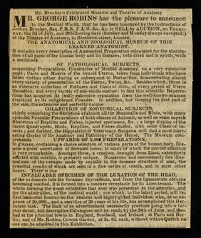
Robins, George Henry, 1777-1847 (1)
Shepherd, Thomas H. (Thomas Hosmer) (1)
Storie, S., active 1780 (1)
Zumbo, Gaetano Giulio, 1656-1701 (1)
Submit
23 results
Search result sorting
Sort by:
Relevance
Production dates
Sort order:
Ascending
Descending
Submit
Page
1
of 1
Close modal window
Page
1
of 1