63 results filtered with: Projections

- Digital Images
- Online
Osteocyte in cortical bone, SEM
Kevin Mackenzie, University of Aberdeen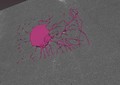
- Digital Images
- Online
Osteocyte in cortical bone, SEM
Kevin Mackenzie, University of Aberdeen
- Digital Images
- Online
A single enteropathogenic E. coli in the intestine
S. Schuller
- Digital Images
- Online
Lung cancer cell.
Anne Weston, Francis Crick Institute
- Digital Images
- Online
Rat neurones, SEM
Anne Weston, Francis Crick Institute
- Digital Images
- Online
Neurone development, embryoid body
John Grady, Doug Turnbull, Claudia Racca, Newcastle University
- Digital Images
- Online
Neurone development, embryoid body
John Grady, Doug Turnbull, Claudia Racca, Newcastle University
- Digital Images
- Online
Human brain cancer stem cells treated with graphene, SEM
Izzat Suffian, Pedro Costa, Stephen Pollard, David McCarthy & Khuloud T. Al-Jamal
- Digital Images
- Online
Bipolar neurone in the midbrain of an adult zebrafish, LM
Dr Mónica Folgueira
- Digital Images
- Online
Human kidney cell, Gated-STED microscopy
Alison Dun, ESRIC (Edinburgh Super-Resolution Imaging Consortium)
- Digital Images
- Online
Lung cancer cell.
Anne Weston, Francis Crick Institute
- Digital Images
- Online
Single neurone in the midbrain of an adult zebrafish, LM
Dr Mónica Folgueira
- Digital Images
- Online
Skin cancer cell
Annie Cavanagh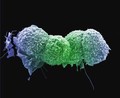
- Digital Images
- Online
Lung cancer cells.
Anne Weston, Francis Crick Institute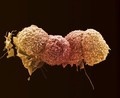
- Digital Images
- Online
Lung cancer cells.
Anne Weston, Francis Crick Institute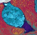
- Digital Images
- Online
Goblet cells are packed full of mucous globules (blue), which they release to provide lubrication and protection to the inner surfaces of the intestine and the respiratory system among others. The mucous globules are condensed inside the goblet cell but expand hugely once they are released, absorbing water within 20 milliseconds. This rapid release occurs in response to lots of different stimuli and allows the mucous to get to work instantly.
University of Edinburgh
- Digital Images
- Online
Lung cancer cell.
Anne Weston, Francis Crick Institute
- Digital Images
- Online
Human small intestine showing villi and glands. The cytokeratinin the cells is stained blue, the cell nuclei are stained red and the endothelial cells lining the blood vessels are stained green.
S. Schuller
- Digital Images
- Online
Rat neurones, SEM
Anne Weston, Francis Crick Institute
- Digital Images
- Online
Lung cancer cells.
Anne Weston, Francis Crick Institute
- Digital Images
- Online
Human small intestine showing villi. The cytokeratinin the cells is stained blue, the cell nuclei are stained red and the endothelial cells lining the blood vessels are stained green.
S. Schuller
- Digital Images
- Online
Mouse colon infected with Citrobacter rodentium
S. Schuller
- Digital Images
- Online
Lung cancer cells.
Anne Weston, Francis Crick Institute
- Digital Images
- Online
Human oral squamous cell carcinoma cell, SEM
Anne Weston, LRI, CRUK and John Marshall, Tumour Biology Lab
- Digital Images
- Online
Neuroendocrine cells in the small intestine
S. Schuller