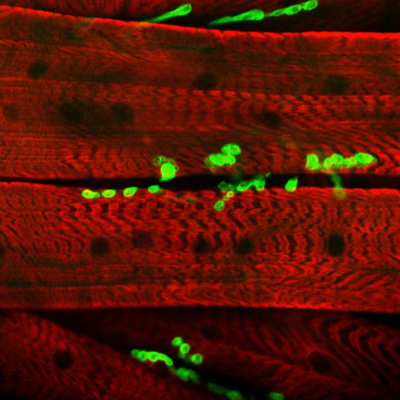
Neuromuscular junction showing the association of the nerves and the muscle fibres. This image is taken from a Drosophila larva and shows the neuromuscular junctions stained with green fluorescence protein (GFP) in association with the muscle fibres in red. The muscle is stained with an antibody which provides the red colour and also shows the striated pattern of the contractile filaments (sarcomers) of the muscle.
- Hermann Aberle, University of Munster
Licence: Attribution 4.0 International (CC BY 4.0)
Credit: Neuromuscular junction showing the association of the nerves and the muscle fibres. This image is taken from a Drosophila larva and shows the neuromuscular junctions stained with green fluorescence protein (GFP) in association with the muscle fibres in red. The muscle is stained with an antibody which provides the red colour and also shows the striated pattern of the contractile filaments (sarcomers) of the muscle. Hermann Aberle, University of Munster. Source: Wellcome Collection.