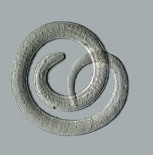
This is a high resolution image of the human helminth parasite Trichinella spiralis. In this image cysts of the parasite have been digested and a single worm isolated and mounted.
The image was created by stacking and stitching together 6 individual images at X25 magnification. Each image in the six image stitch was in turn produced by stacking individual images at different levels of focus through the subject. Altogether 60 individual photographs were used to create this image. The imaging technique used was differential interference contrast (DIC).
Trichinella are the smallest nematode parasite of humans. Humans are infected with the parasite following ingestion of infected meat, such as pork. Adult worms produce larvae in the intestine of the host, which bore through the intestinal wall into the blood and lymphatic system. The parasites are then carried to striated muscle where they become enclosed in a capsule or cyst. The boring of the worms through the intestinal wall produces an immunological respose resulting in symptoms such as nausea, sweating and diarrhea. Around ten days post-infection, more severe symptoms occur including muscle pain, weak pulse and blood pressure, heart and nerve damage, eventually leading to death from heart failure, respiratory complications or kidney malfunction.