Progressive multifocal leukoencephalopathy (PML)
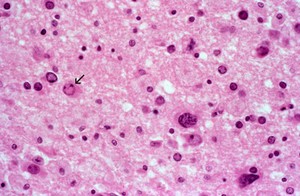
This is a high power microscopic view of a section of white matter from the brain of an adult with PML, which shows a large astrocyte near the centre. Many of the other small cells have eosinophilic, pink-staining, intranuclear inclusions which are collections of JC virus, the causative agent of PML. The arrow indicates an infected oligodendrocyte. H&E stain.
The infected cells are oligodendrocytes, which make myelin. Virus infection and damage to the oligodendrocytes results in demyelination. Inclusions are best seen in areas of white matter that are less damaged (ie. in the early phase of disease).