Despite our apparent perfect bilateral symmetric body plan, we all show small but conspicuous left-right asymmetries that are thought to be evolutionarily advantageous. Brain lateralisation, for instance, leads to an increase in cognitive performance. Zebrafish are useful for studying how left-right brain asymmetries arise during embryonic development and how the resultant asymmetric neuronal circuitry ultimately impacts on function and behaviour.
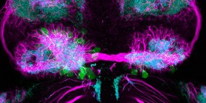
This image shows the habenular nucleus of a zebrafish embryo 4 days post-fertilization. The habenular nucleus is a part of the epithalamus which connects the limbic system (responsible for instinct and mood) to other regions of the brain.
The arrangement of the habenular axons is highlighted in magenta in this image, and the synaptic neuropil in cyan. Neurons expressing the gene left-over (lov) are highlighted in green, and are predominantly enriched in the left habenula. Different neuronal subtypes, with a typical molecular signature and neural connectivity pattern, are produced in different ratios in the left and right habenular nucleus and confer their unique asymmetric nature.
Width of image is 200 micrometres.