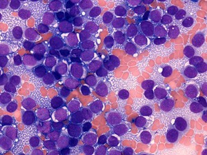
Direct smear stained with MGG (May Grunwald Giemsa) prepared from fine needle biopsy of paravertebral mass in a 20 year old man
Peripheral Neuroectodermal Tumour is a rare form of cancer that tends to occur in young people. Morphologically is characterized by a monotonous population of relatively small round tumour cells, a pattern also seen in other tumours such as lymphoma, rhabdomyosarcoma and neuroblastoma. Collectively tumours with this appearance are often referred to as "small round blue cell tumours". A important clue to the diagnosis of PNET in cytology is that the tumour cells, unlike those of lymphoma, accumulate glycogen in their cytoplasm. In air dried smears this glycogen rich cytosol forms a reticulated background referred to as "tigroid" identical to that seen in other glycogen rich tumours such as seminoma. The diagnosis of PNET is confirmed by immunohistochemical and cytogenetic analysis to confirm the presence of specific translocations, usually between chromosome 11 and 22.
This image shows a population of round tumour cells with purple nuclei and small light blue nucleoli, orange red blood cells and a pale grey blue reticulated tigroid background.
Image width 215 microns